Peripheral Blood Smear
Peripheral Blood Smear
HematologyWright-stained peripheral blood film showing red blood cells, white blood cells, and platelets
Drag & drop images here to add to this specimen
or click Upload Image aboveRelease to upload
Image Library
28 images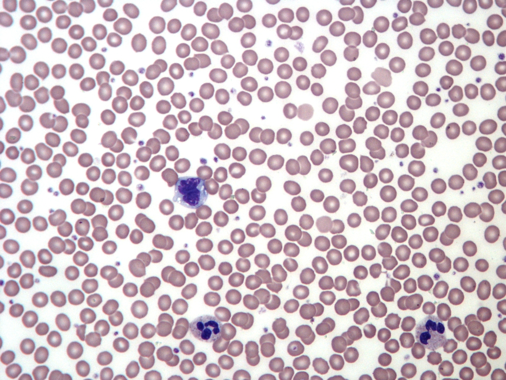
Normal peripheral blood smear showing biconcave red blood cells, scattered platelets, and a segmented neutrophil. Classic Wright-stained morphology.
Oil immersion, ideal reference for normal hematology
Wikimedia Commons — CC BY-SA 3.0
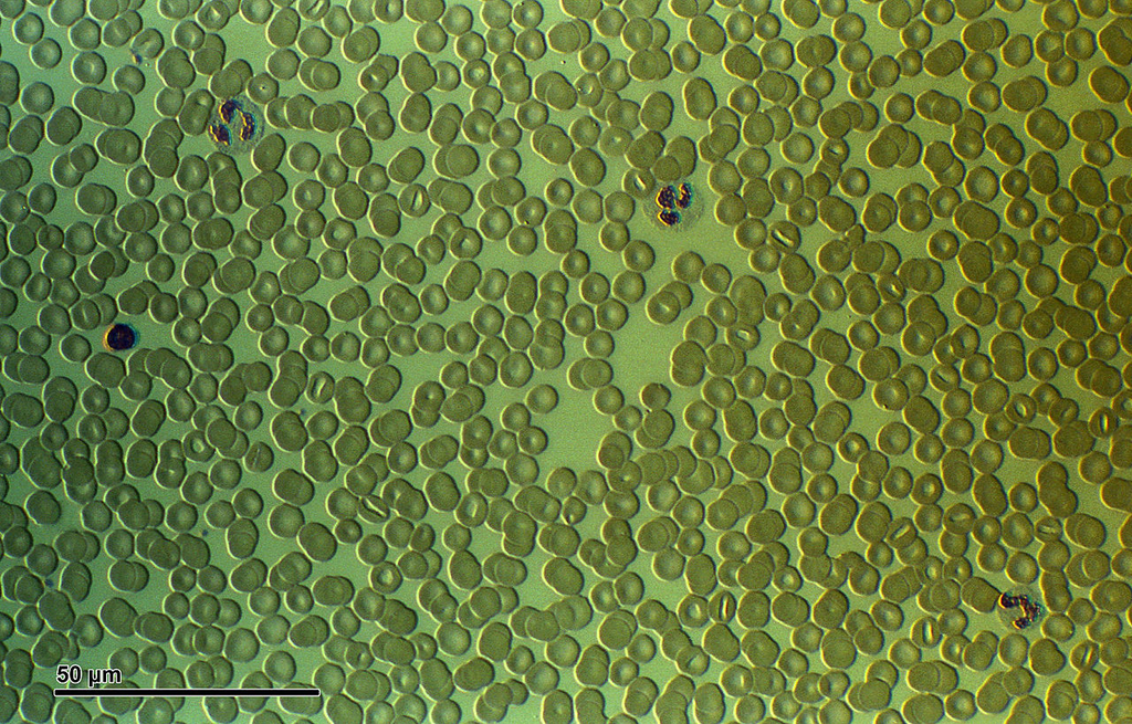
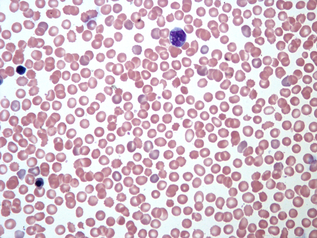
High-resolution photomicrograph of human blood smear showing detailed red blood cell morphology and scattered leukocytes. Excellent for morphological assessment.
High resolution image suitable for detailed cell morphology analysis
Wikimedia Commons — CC BY-SA 4.0
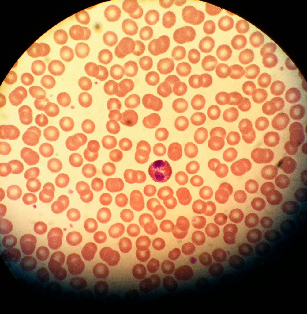
Wright-stained smear showing an eosinophil with characteristic bilobed nucleus and bright red-orange granules, surrounded by normal red blood cells and platelet clusters.
Oil immersion — eosinophil identification reference
Wikimedia Commons — Public Domain
Clinical blood cell microscopy image showing red blood cells and white blood cells. U.S. government public domain image used for medical training.
U.S. Air Force medical training image
Wikimedia Commons — Public Domain (U.S. Government)

Diagnostic-quality peripheral blood smear with well-distributed cells. Multiple white blood cell types visible among normal red blood cells.
Diagnostic quality, feathered edge preparation
Wikimedia Commons — CC BY-SA 4.0
Blood smear highlighting monocytes — large white blood cells with kidney-shaped nuclei and abundant gray-blue cytoplasm. Surrounded by normal erythrocytes.
Oil immersion — monocyte identification and morphology reference
Wikimedia Commons — CC BY-SA 3.0
Peripheral blood smear from a neonate showing larger red blood cells (macrocytes) typical of newborn hematology. Nucleated red blood cells may be present.
Neonatal specimen — larger RBCs and possible nucleated cells are normal findings
Wikimedia Commons — CC BY-SA 3.0
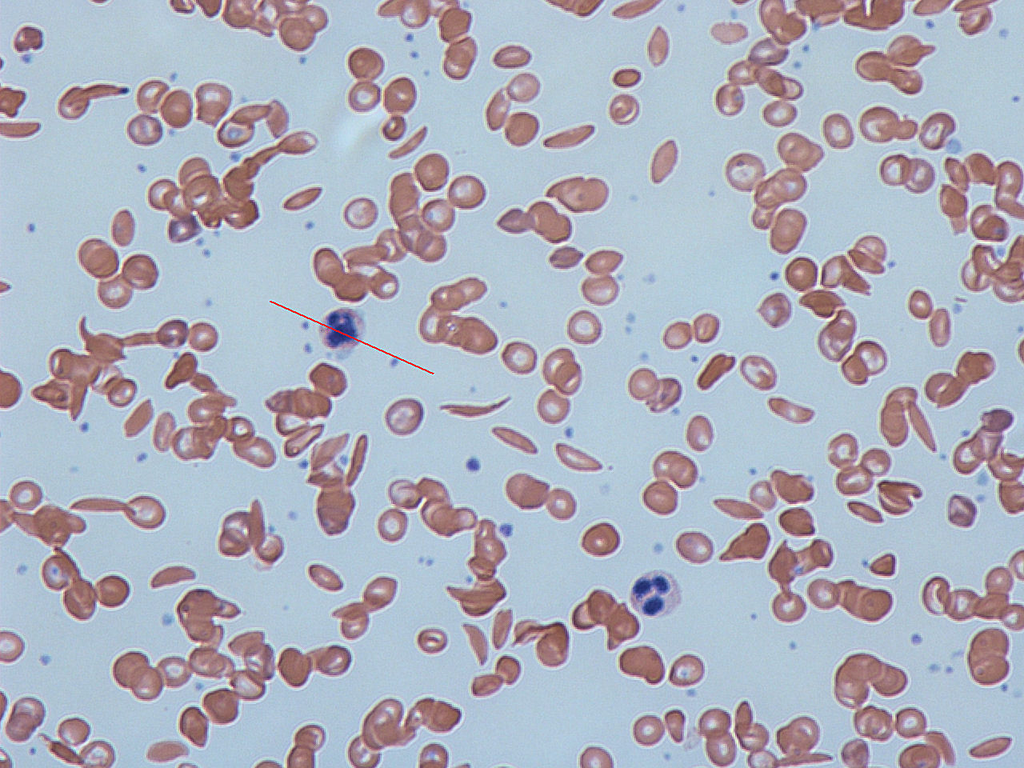
Blood smear from a patient with sickle cell disease showing characteristic sickled erythrocytes (drepanocytes). Sickled cells are elongated, crescent-shaped red blood cells caused by abnormal hemoglobin S polymerization.
Sickle cell anemia — HbSS disease, classic sickled erythrocyte morphology
Wikimedia Commons — CC BY-SA 3.0
Reference micrograph of sickle-shaped red blood cells from the National Institute of Diabetes and Digestive and Kidney Diseases. Shows classic drepanocyte morphology.
NIH/NIDDK reference image for sickle cell disease education
Wikimedia Commons — Public Domain (US Gov, NIH/NIDDK)
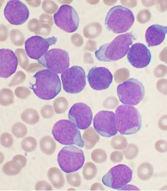
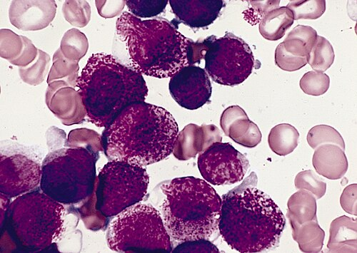
Wright-stained bone marrow aspirate smear from a patient with precursor B-cell acute lymphoblastic leukemia. Numerous lymphoblasts with scant cytoplasm, fine chromatin, and indistinct nucleoli are visible.
ALL — most common childhood leukemia, blast cells predominate in marrow
Wikimedia Commons — CC BY-SA 3.0
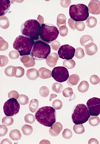
Bone marrow aspirate from a 3-year-old male diagnosed with acute lymphoblastic leukemia, FAB L1 subtype. Small uniform blasts with high nuclear-to-cytoplasmic ratio and inconspicuous nucleoli.
ALL-L1 (FAB classification) — small uniform blasts, favorable prognosis subtype
Wikimedia Commons — Public Domain (US Gov, AFIP)
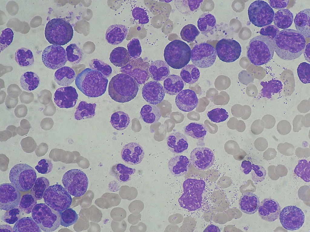
CML blood smear showing marked leukocytosis with granulocyte left shift. Myeloid cells at various stages of maturation are visible, including myelocytes, metamyelocytes, bands, and segmented neutrophils.
CML — Philadelphia chromosome positive, BCR-ABL1 fusion, granulocyte left shift
Wikimedia Commons — CC BY-SA 3.0
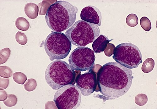
Bone marrow aspirate showing acute myeloid leukemia M1 subtype with agranular myeloblasts. Blasts have fine chromatin, prominent nucleoli, and scant cytoplasm without significant granulation.
AML-M1 (FAB) — minimally differentiated, >90% blasts in marrow
Wikimedia Commons — Public Domain (US Gov, AFIP)
Bone marrow aspirate from hypergranular acute promyelocytic leukemia (APL). Promyelocytes with abundant azurophilic granulation and characteristic Auer rods are visible. APL is associated with t(15;17) translocation.
AML-M3/APL — Auer rods present, responds to all-trans retinoic acid (ATRA)
Wikimedia Commons — Public Domain (US Gov, AFIP)
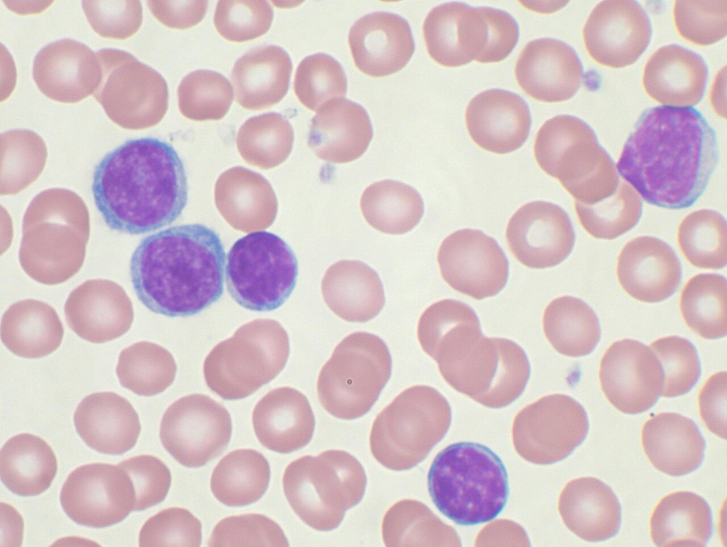
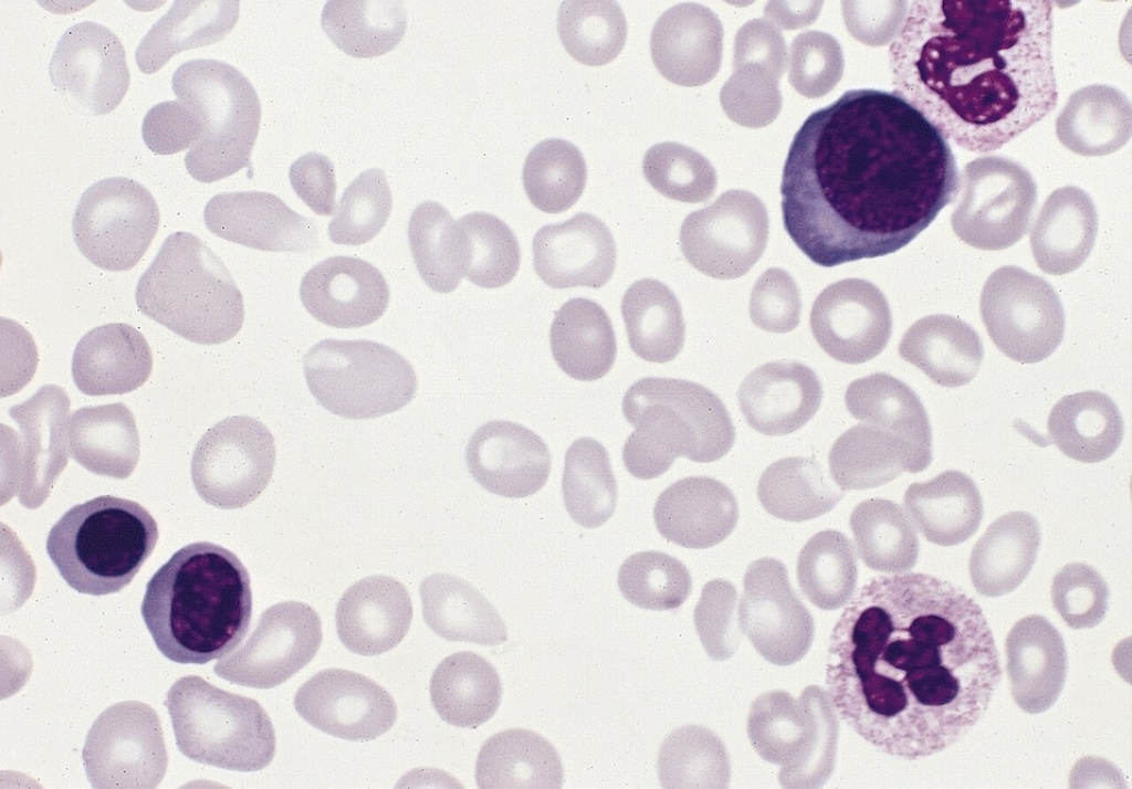
CLL peripheral blood smear showing abundant small mature lymphocytes with condensed chromatin and scant cytoplasm. Smudge cells (basket cells) are characteristically present, representing fragile neoplastic lymphocytes.
CLL — most common adult leukemia, smudge cells are hallmark finding
Wikimedia Commons — CC BY-SA 3.0
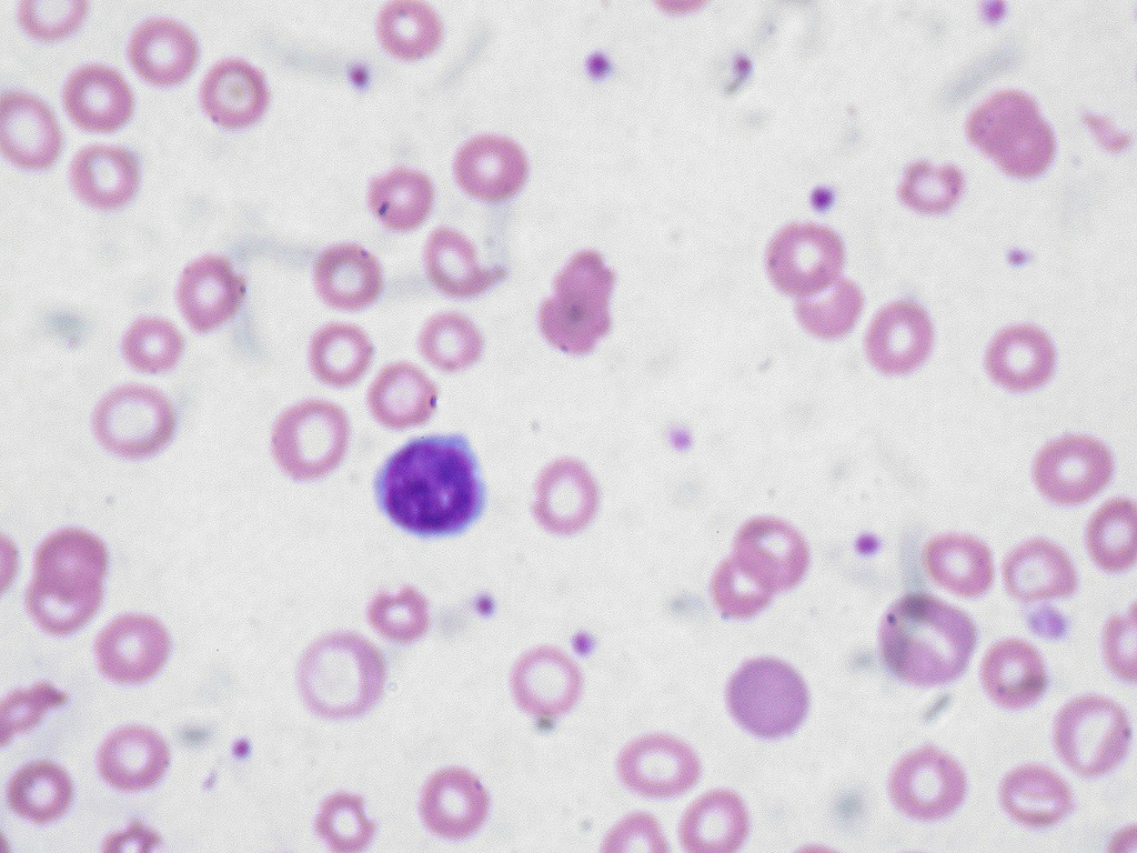
Peripheral blood film showing hypochromic microcytic red blood cells characteristic of iron deficiency anemia. Cells show increased central pallor due to reduced hemoglobin content and are smaller than normal.
Iron deficiency — most common cause of anemia worldwide, low MCV and MCH
Wikimedia Commons — CC BY-SA 2.0
Detailed blood film demonstrating iron deficiency anemia with microcytic hypochromic erythrocytes. Target cells and pencil cells (elliptocytes) may also be present. Anisocytosis and poikilocytosis are evident.
Iron deficiency — anisocytosis, poikilocytosis, elevated RDW on CBC
Wikimedia Commons — CC BY-SA 3.0
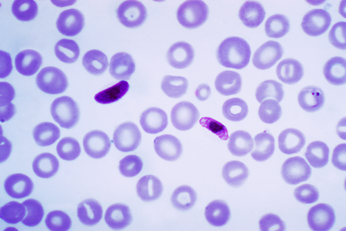
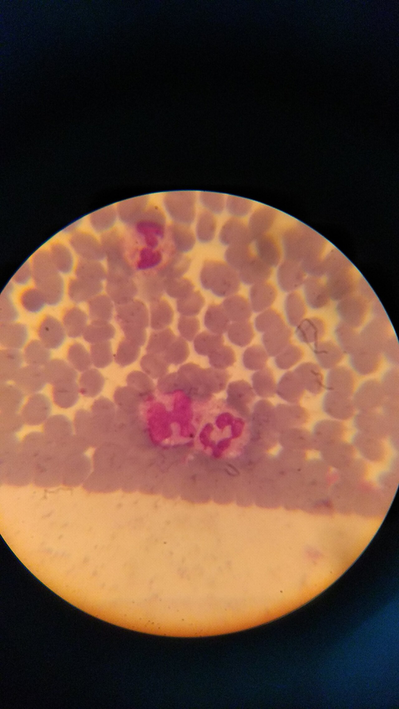
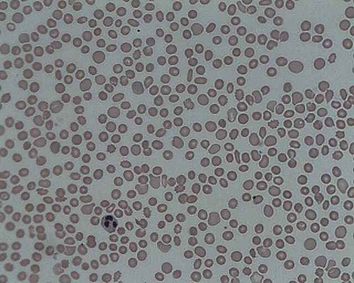
Blood smear showing Plasmodium falciparum gametocytes among red blood cells. The characteristic banana/crescent-shaped gametocytes are pathognomonic for P. falciparum malaria, the most severe form of malaria.
P. falciparum malaria — banana-shaped gametocytes, CDC reference
Wikimedia Commons — Public Domain (US CDC)
Micrograph of plasmacytoma showing abundant malignant plasma cells with eccentric nuclei and perinuclear hof (pale zone). Occasional Mott cells with intracytoplasmic Russell bodies (immunoglobulin inclusions) are visible.
Multiple myeloma / plasmacytoma — clonal plasma cell neoplasm, Mott cells present
Wikimedia Commons — CC BY-SA 3.0
Blood smear from a 68-year-old woman with a 13-year history of polycythemia vera. Shows increased red blood cell precursors and anisopoikilocytosis. PV is a myeloproliferative neoplasm with JAK2 V617F mutation.
Polycythemia vera — JAK2-positive MPN, elevated RBC mass, risk of thrombosis
Wikimedia Commons — Public Domain (US Gov, AFIP)
Blood film showing spherocytes — abnormally round, dense red blood cells lacking the normal biconcave shape. Spherocytes have reduced surface-to-volume ratio and are prone to splenic sequestration and hemolysis.
Hereditary spherocytosis — membrane protein defect, osmotic fragility increased
Wikimedia Commons — CC BY-SA 4.0
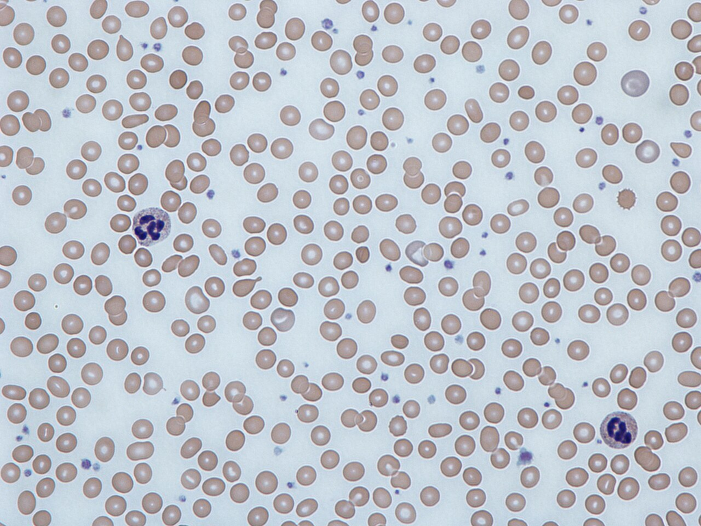
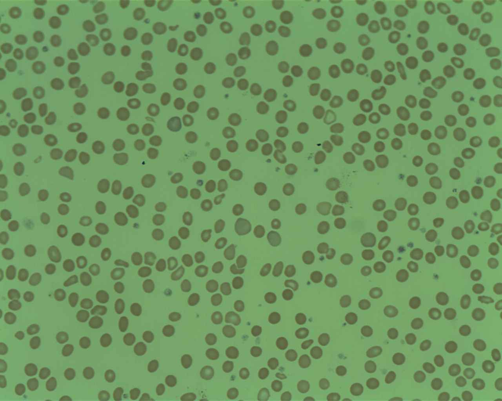
Blood film from a thrombocytopenic patient showing a near-absence of platelets. Normal blood films show 7-21 platelets per oil-immersion field; this field shows virtually none, consistent with severe thrombocytopenia.
Severe thrombocytopenia — <20,000/μL, risk of spontaneous bleeding
Wikimedia Commons — CC BY-SA 4.0
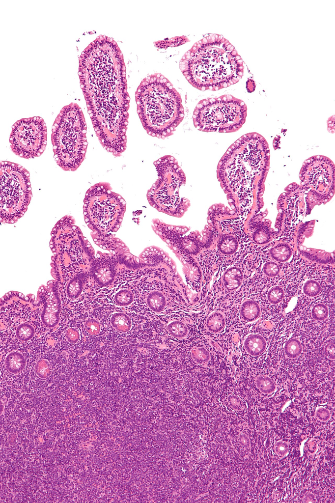
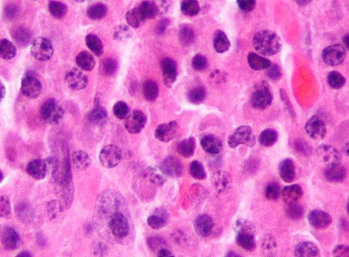
Intermediate magnification micrograph of mantle cell lymphoma of the terminal ileum. Monomorphic small-to-medium lymphoid cells with irregular nuclear contours and abundant mitotic figures are characteristic.
Mantle cell lymphoma — cyclin D1 positive, t(11;14), aggressive B-cell NHL
Wikimedia Commons — CC BY-SA 3.0
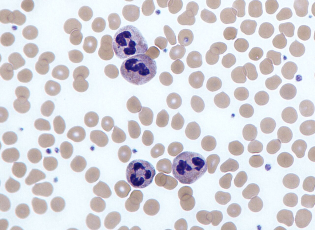
Four neutrophil white blood cells at high magnification showing multilobed (segmented) nuclei with fine chromatin and pale pink cytoplasm with fine granules. Surrounded by erythrocytes and scattered platelets.
Neutrophils — most abundant WBC type, 3-5 nuclear lobes, first responders to infection
Wikimedia Commons — CC BY-SA 3.0
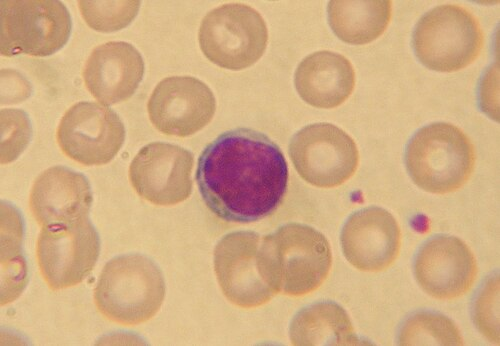
Single lymphocyte in a human blood smear at oil-immersion magnification. Shows a large round nucleus with dense chromatin occupying most of the cell, surrounded by a thin rim of basophilic cytoplasm.
Small lymphocyte — T or B cell, dense nuclear chromatin, high N:C ratio
Wikimedia Commons — CC BY-SA 3.0
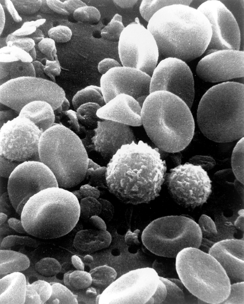
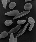
Scanning electron micrograph of normal circulating human blood showing the 3D surface morphology of erythrocytes (biconcave discs), leukocytes (rough surface), and platelets (small irregular fragments).
SEM — NCI reference, shows true 3D morphology of blood cell types
Wikimedia Commons — Public Domain (US Gov, NCI)
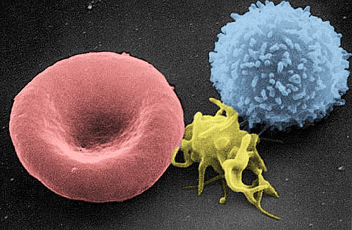
Colorized scanning electron micrograph showing an erythrocyte (red), a thrombocyte/platelet (yellow), and a leukocyte (blue-green). Demonstrates the relative size and surface texture differences between blood cell types.
Colorized SEM — relative size comparison of blood cell types, NCI reference
Wikimedia Commons — Public Domain (US Gov, NCI-Frederick)
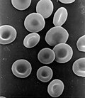
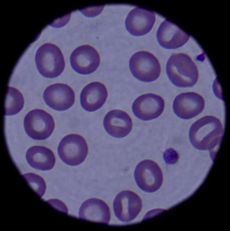
Close-up micrograph of red blood cells (erythrocytes) showing their characteristic biconcave disc shape and uniform size. Reference image for normal RBC morphology.
Normal RBC morphology — 6-8 μm diameter, biconcave disc, no nucleus
Wikimedia Commons — Public Domain (US Gov, NIH/NIDDK)
Compatible Microscopes
| Model | Manufacturer | Type | Magnification Range | NA Max | Resolution |
|---|---|---|---|---|---|
| BZ-X810 | Keyence | Fluorescence All In One | 2–100× | 1.45 | 190 nm |
| VHX-7000 | Keyence | Digital Microscope | 0.1–6000× | — | 100 nm |
| DM6 B | Leica Microsystems | Upright Optical | 1.25–100× | 1.4 | 200 nm |
| DMi8 | Leica Microsystems | Inverted Optical | 2.5–100× | 1.47 | 185 nm |
| Eclipse Ti2 | Nikon | Inverted Optical | 2–100× | 1.45 | 190 nm |
| Eclipse Ni | Nikon | Upright Optical | 2–100× | 1.4 | 200 nm |
| BX53 | Olympus (Evident) | Upright Optical | 2–100× | 1.4 | 200 nm |
| IX83 | Olympus (Evident) | Inverted Optical | 2–100× | 1.4 | 200 nm |
| Axio Observer 7 | Zeiss | Inverted Optical | 5–100× | 1.4 | 200 nm |
| Axio Imager 2 | Zeiss | Upright Optical | 1.25–100× | 1.4 | 200 nm |
| Primostar 3 | Zeiss | Upright Optical | 4–100× | 1.25 | 350 nm |
Edit Image Metadata
AI Generate Image — Peripheral Blood Smear
Fill in the imaging criteria below. A detailed prompt will be built from your selections and sent to OpenAI to generate a realistic microscopy image.