Bone Marrow Aspirate
Bone Marrow Aspirate
HematologyWright-Giemsa stained bone marrow aspirate smear
Drag & drop images here to add to this specimen
or click Upload Image aboveRelease to upload
Image Library
4 images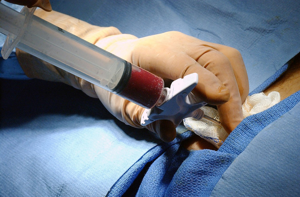
Wright-Giemsa stained bone marrow aspirate smear showing a diverse population of hematopoietic cells at various stages of maturation: myeloid precursors, erythroid precursors, megakaryocytes, and mature blood cells among fat cells.
Bone marrow aspirate — mixed hematopoietic lineages visible
Wikimedia Commons — CC BY-SA 3.0
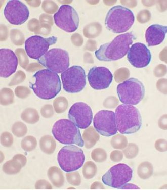
Wright-stained bone marrow aspirate smear showing a predominance of lymphoblasts characteristic of acute lymphoblastic leukemia (ALL). Blasts are medium-sized cells with scant cytoplasm, high nuclear-to-cytoplasmic ratio, and fine chromatin.
ALL — lymphoblasts predominate, high N:C ratio, fine chromatin
Wikimedia Commons — CC BY-SA 3.0
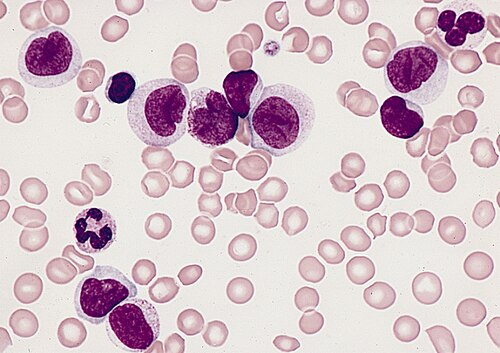
Wright-Giemsa stained bone marrow smear showing acute myelomonocytic leukemia (AML-M4). Both myeloblasts and monocytic precursors are present. Monocytic cells show folded nuclei and gray-blue cytoplasm.
AML-M4 — myeloblasts and promonocytes, mixed lineage
Wikimedia Commons — CC BY-SA 3.0

Wright-Giemsa stained bone marrow aspirate showing abnormal promyelocytes characteristic of acute promyelocytic leukemia (AML-M3). Cells show heavy azurophilic granulation and Auer rods in the cytoplasm.
AML-M3 (APL) — hypergranular promyelocytes, Auer rods, diagnostic morphology
Wikimedia Commons — CC BY-SA 3.0
Compatible Microscopes
| Model | Manufacturer | Type | Magnification Range | NA Max | Resolution |
|---|---|---|---|---|---|
| DM6 B | Leica Microsystems | Upright Optical | 1.25–100× | 1.4 | 200 nm |
| DMi8 | Leica Microsystems | Inverted Optical | 2.5–100× | 1.47 | 185 nm |
| Eclipse Ti2 | Nikon | Inverted Optical | 2–100× | 1.45 | 190 nm |
| Eclipse Ni | Nikon | Upright Optical | 2–100× | 1.4 | 200 nm |
| BX53 | Olympus (Evident) | Upright Optical | 2–100× | 1.4 | 200 nm |
| IX83 | Olympus (Evident) | Inverted Optical | 2–100× | 1.4 | 200 nm |
| Axio Observer 7 | Zeiss | Inverted Optical | 5–100× | 1.4 | 200 nm |
| Axio Imager 2 | Zeiss | Upright Optical | 1.25–100× | 1.4 | 200 nm |
| Primostar 3 | Zeiss | Upright Optical | 4–100× | 1.25 | 350 nm |
Edit Image Metadata
AI Generate Image — Bone Marrow Aspirate
Fill in the imaging criteria below. A detailed prompt will be built from your selections and sent to OpenAI to generate a realistic microscopy image.