Specimen Image Library
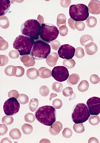
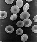
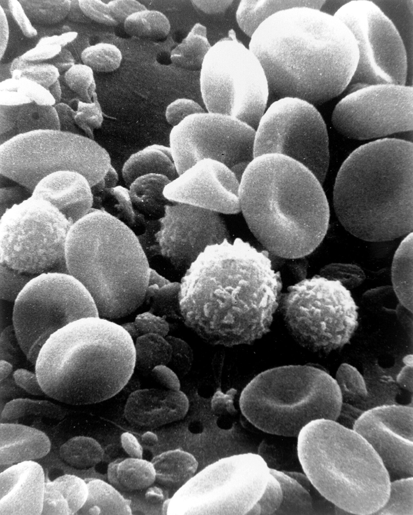
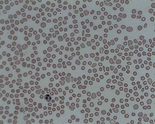
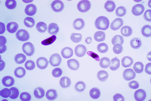
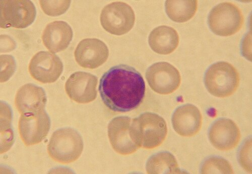
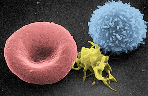
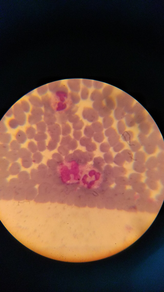
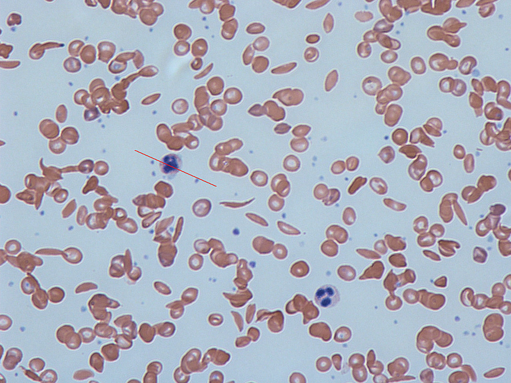

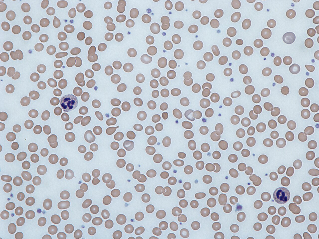
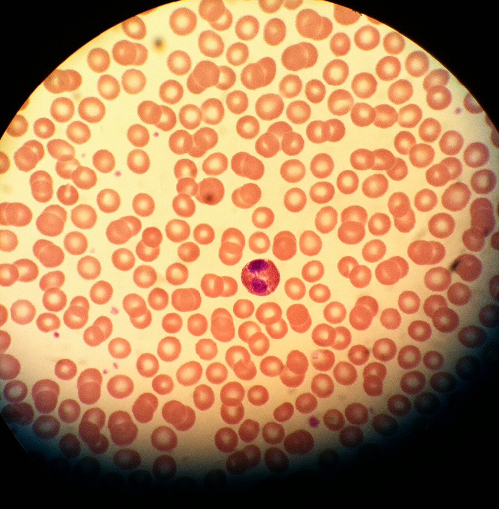
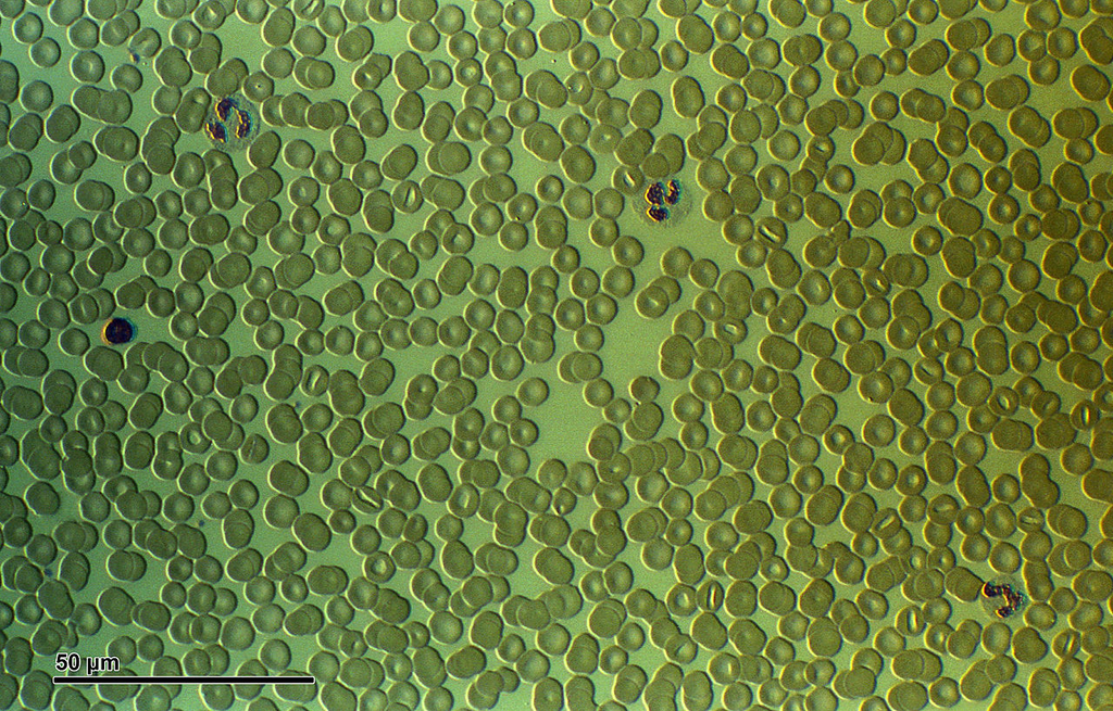

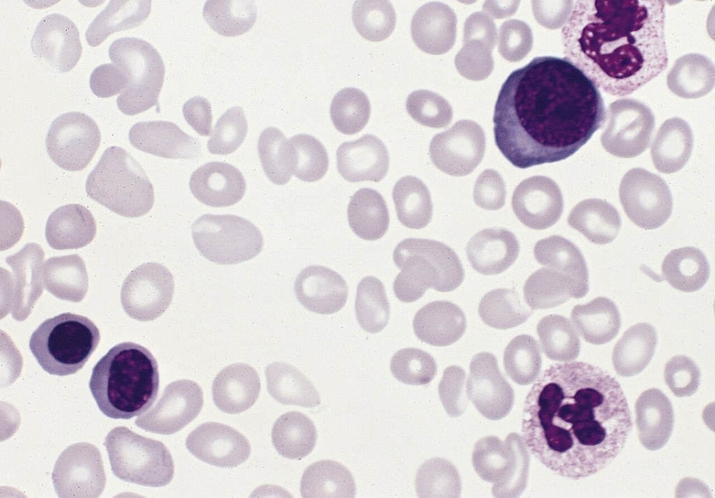
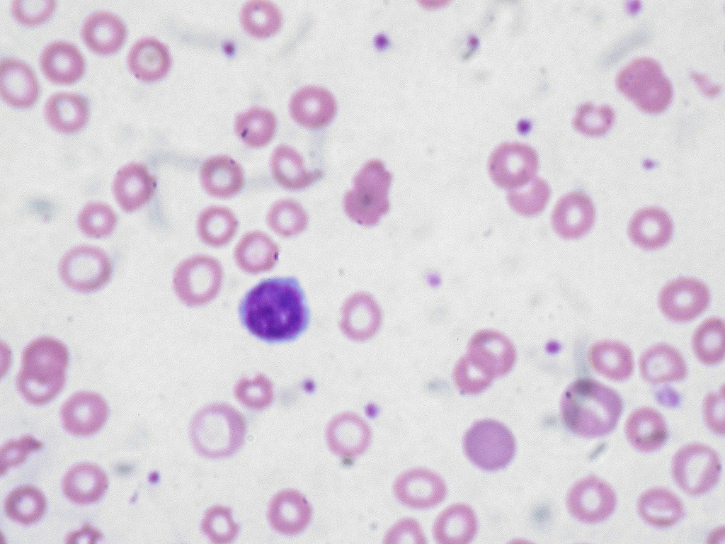
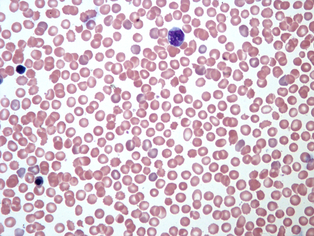
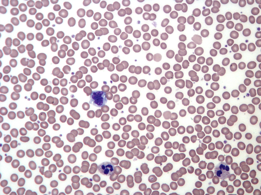
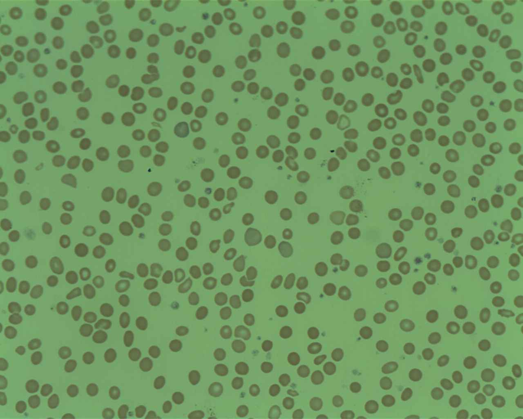
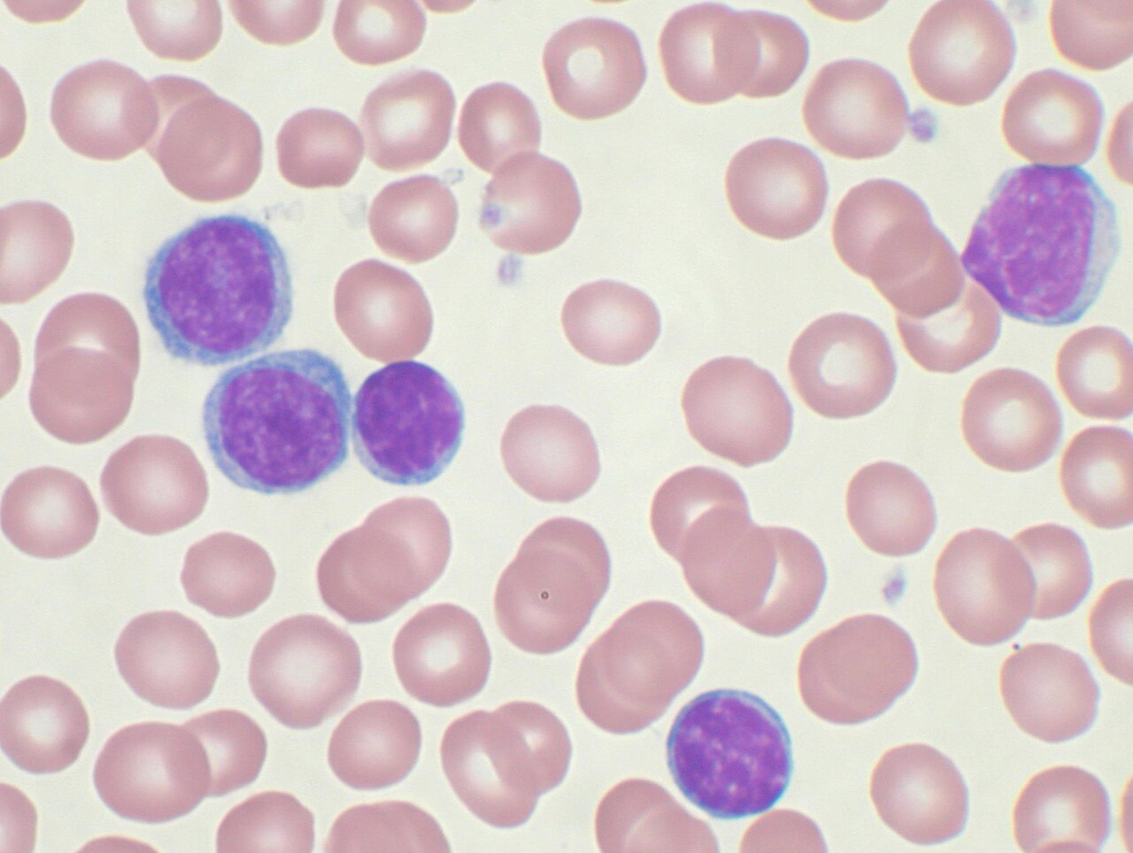
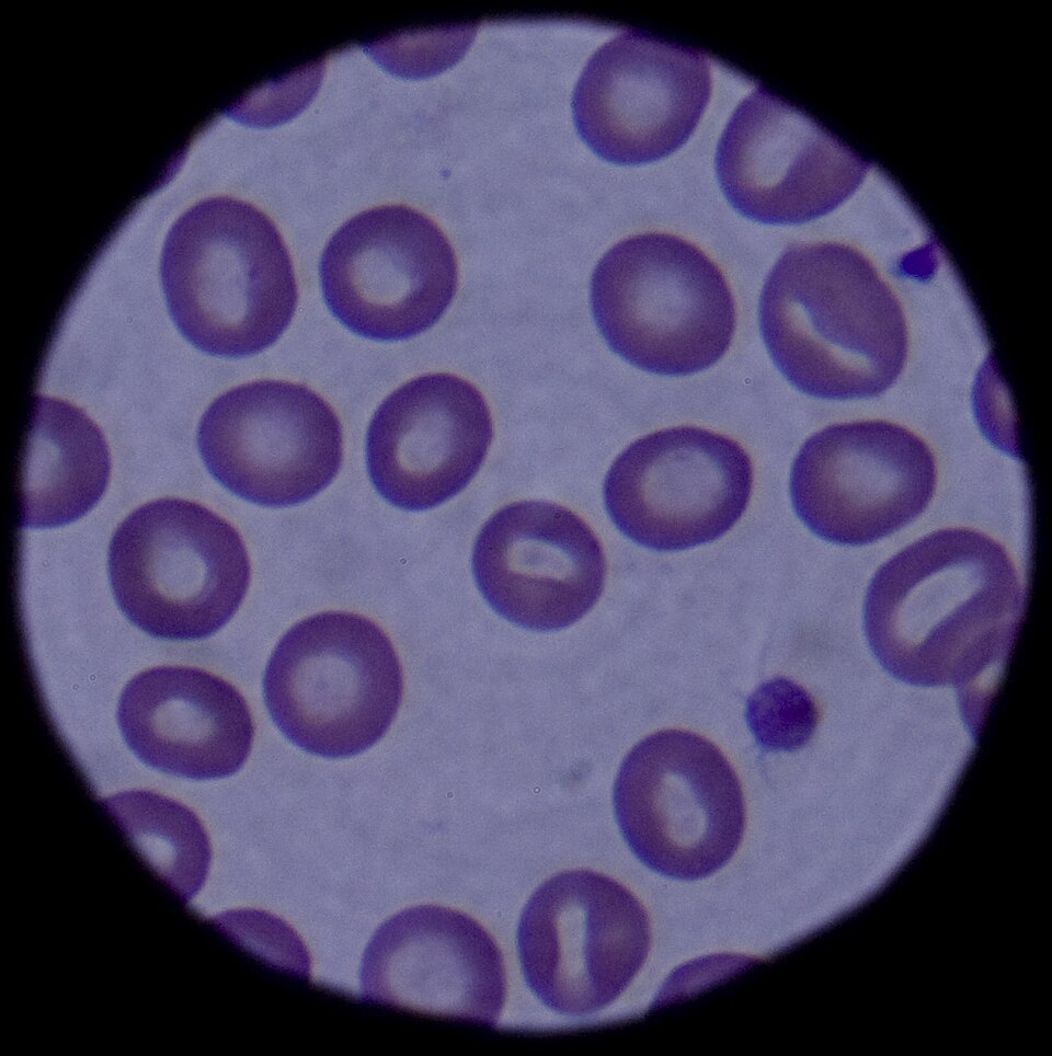
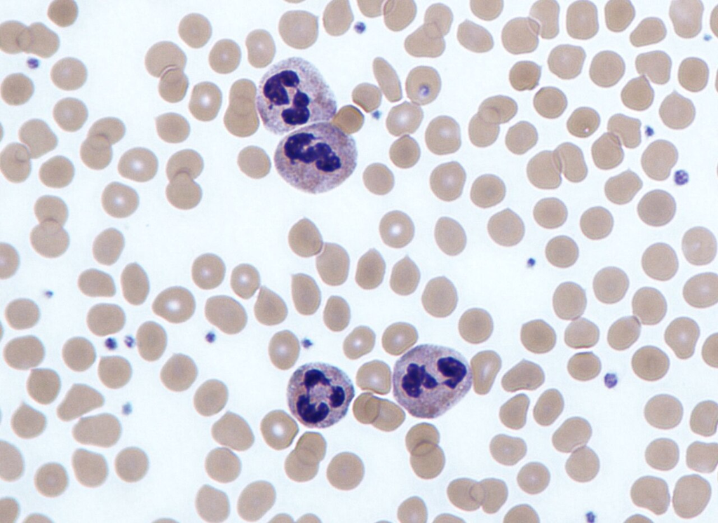

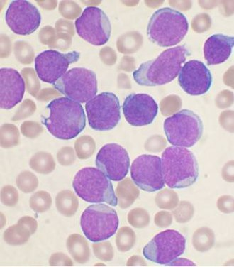
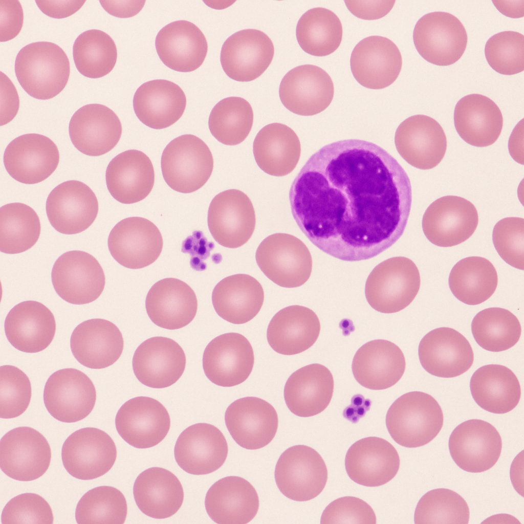
Wright-stained peripheral blood film showing red blood cells, white blood cells, and platelets
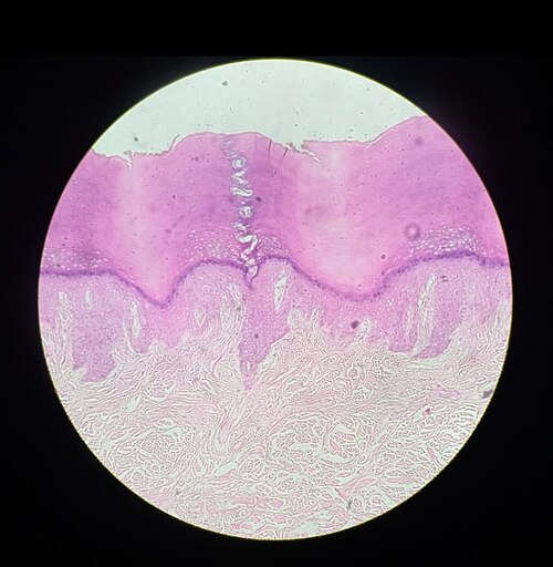
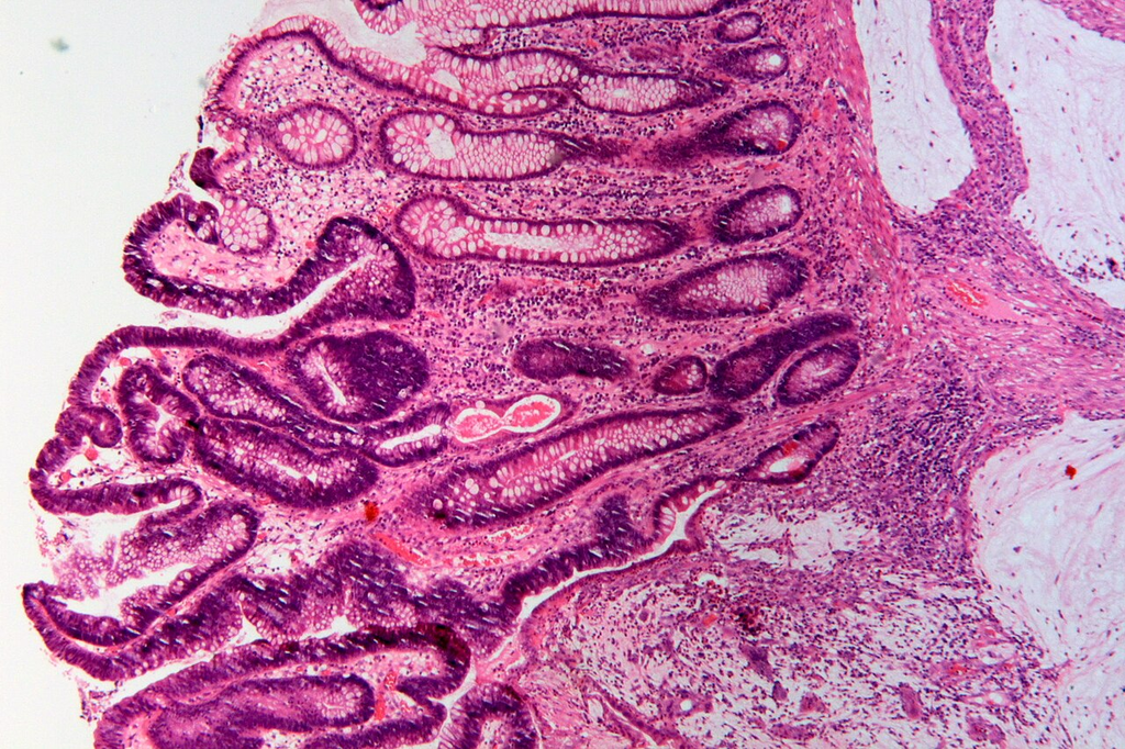
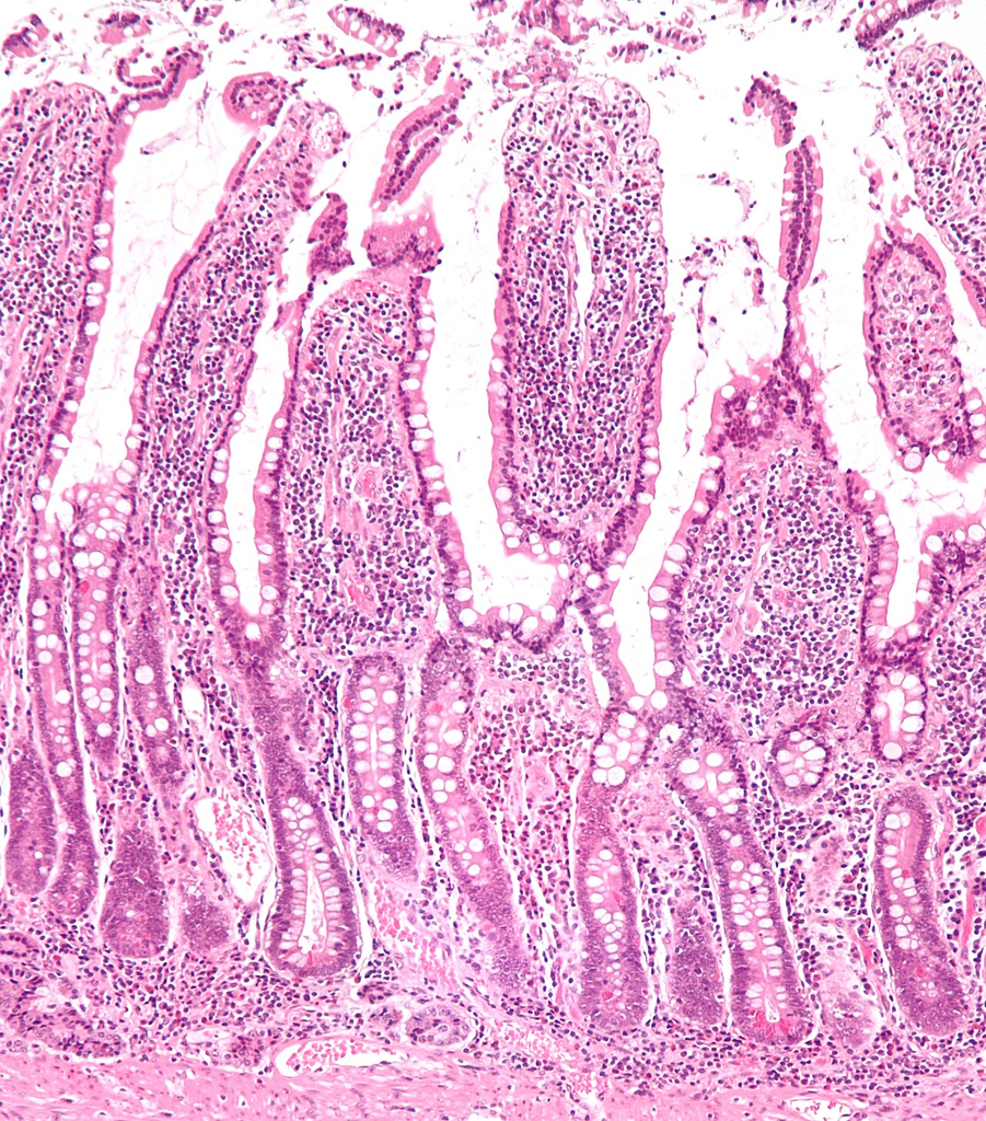
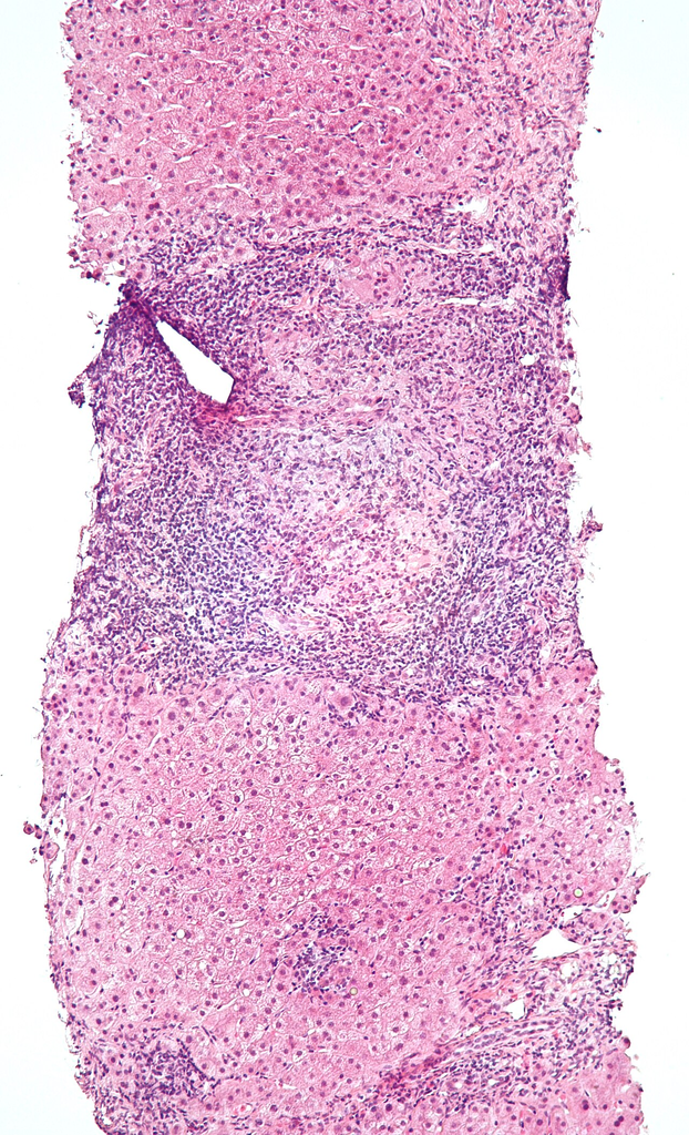
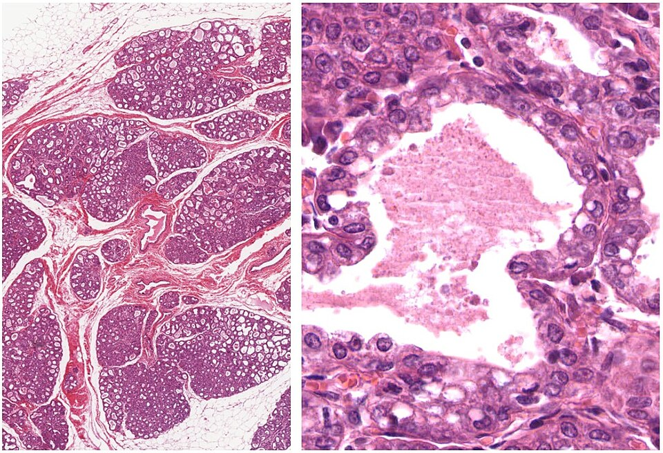
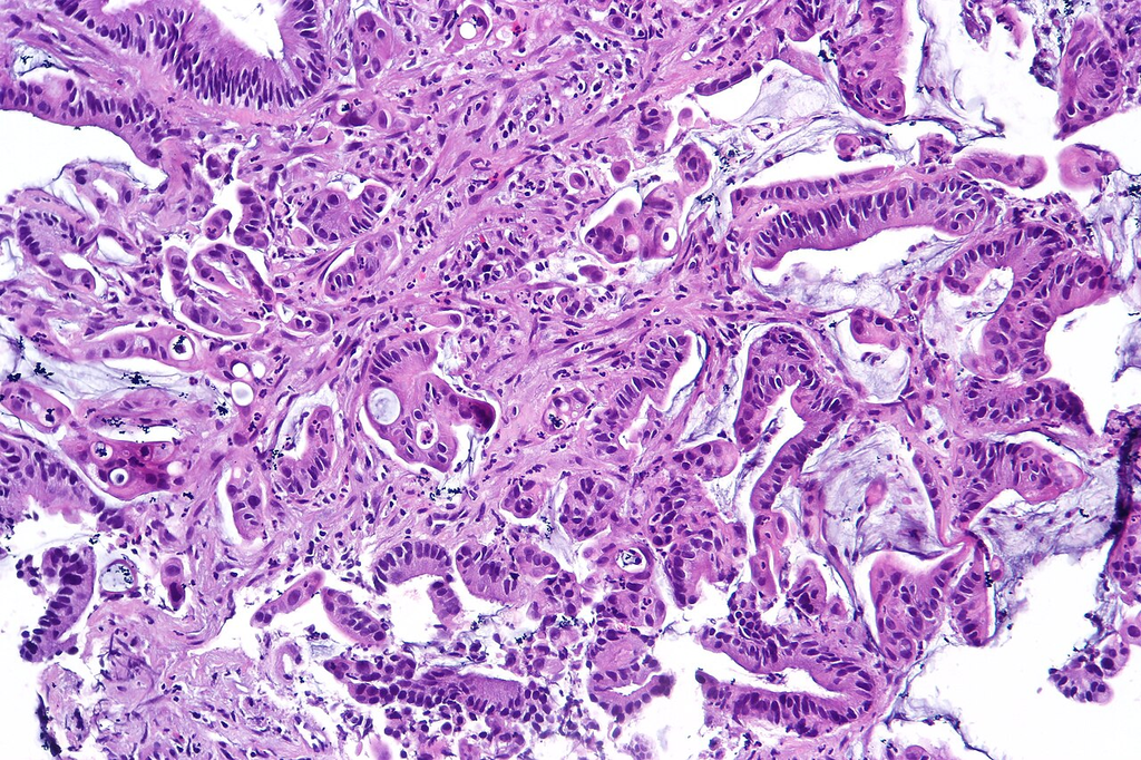
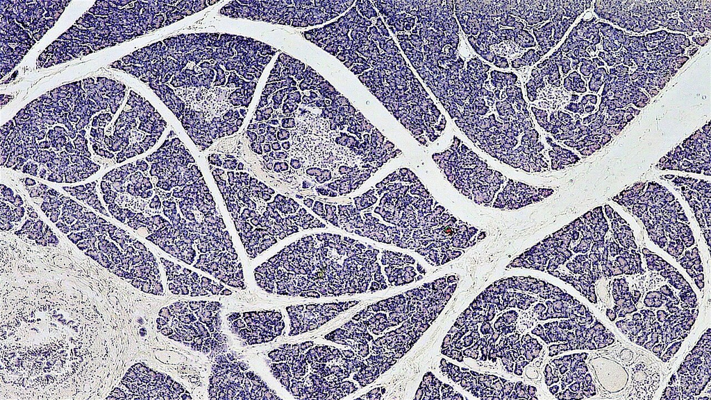
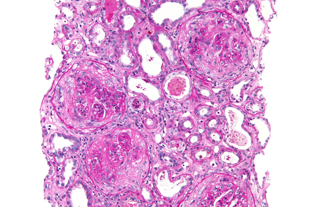
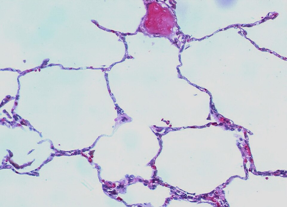
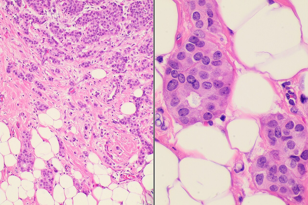
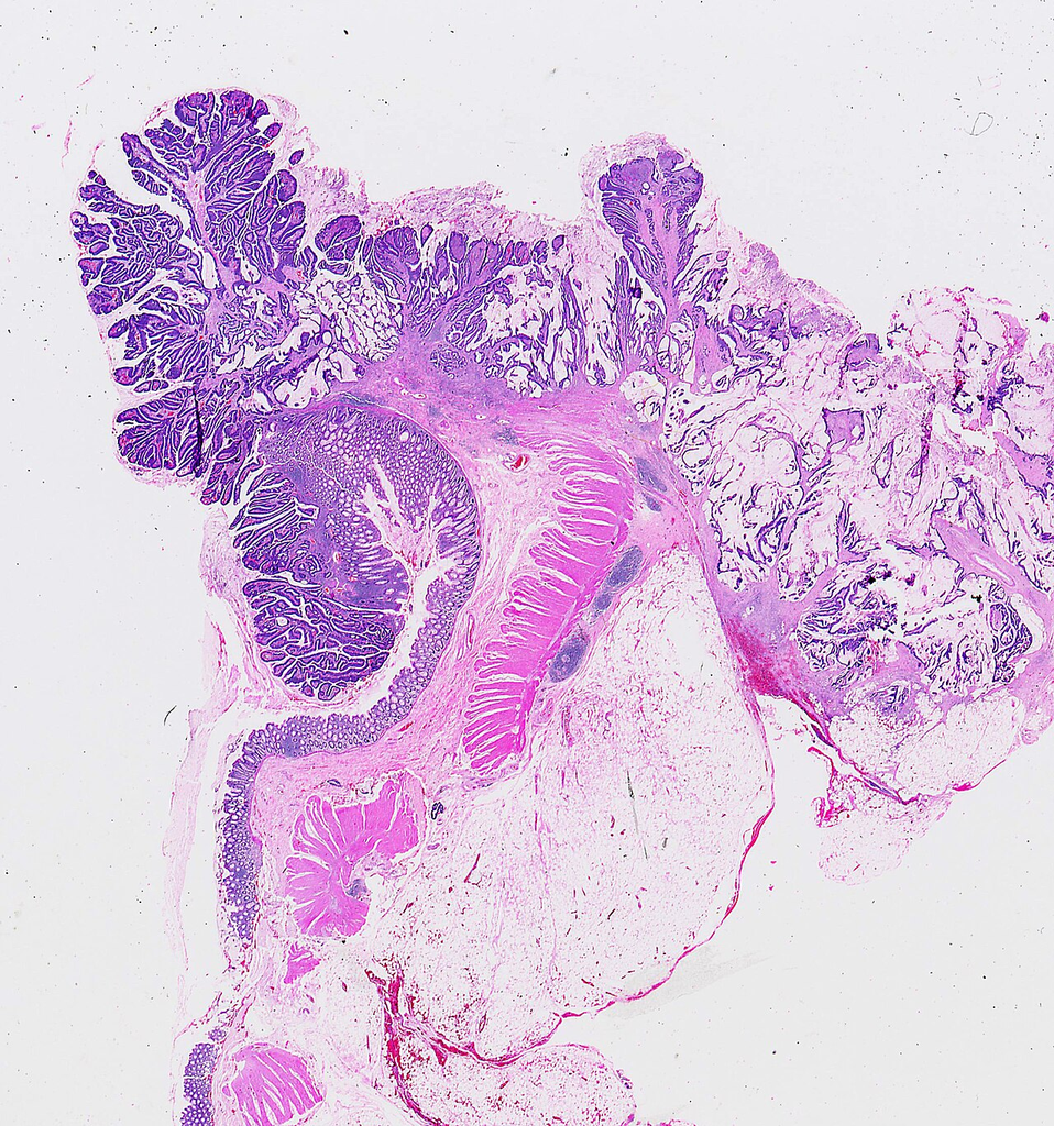
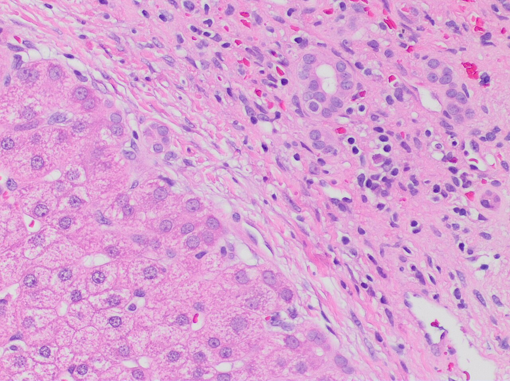
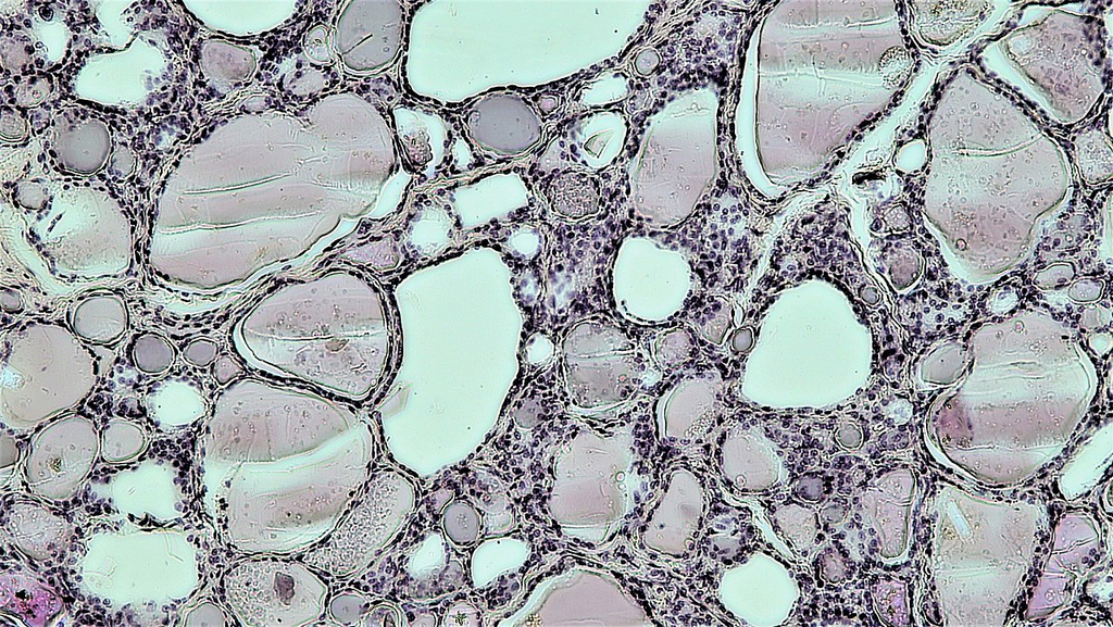
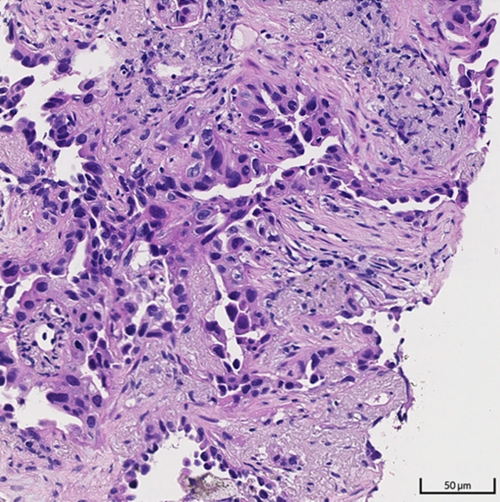
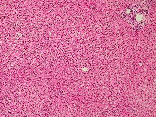
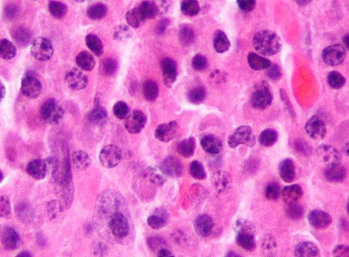
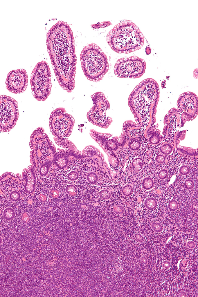
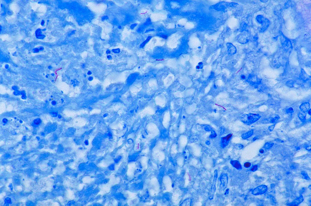
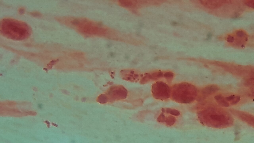
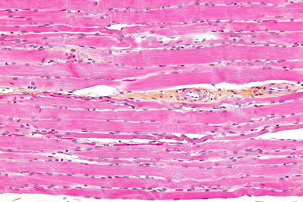
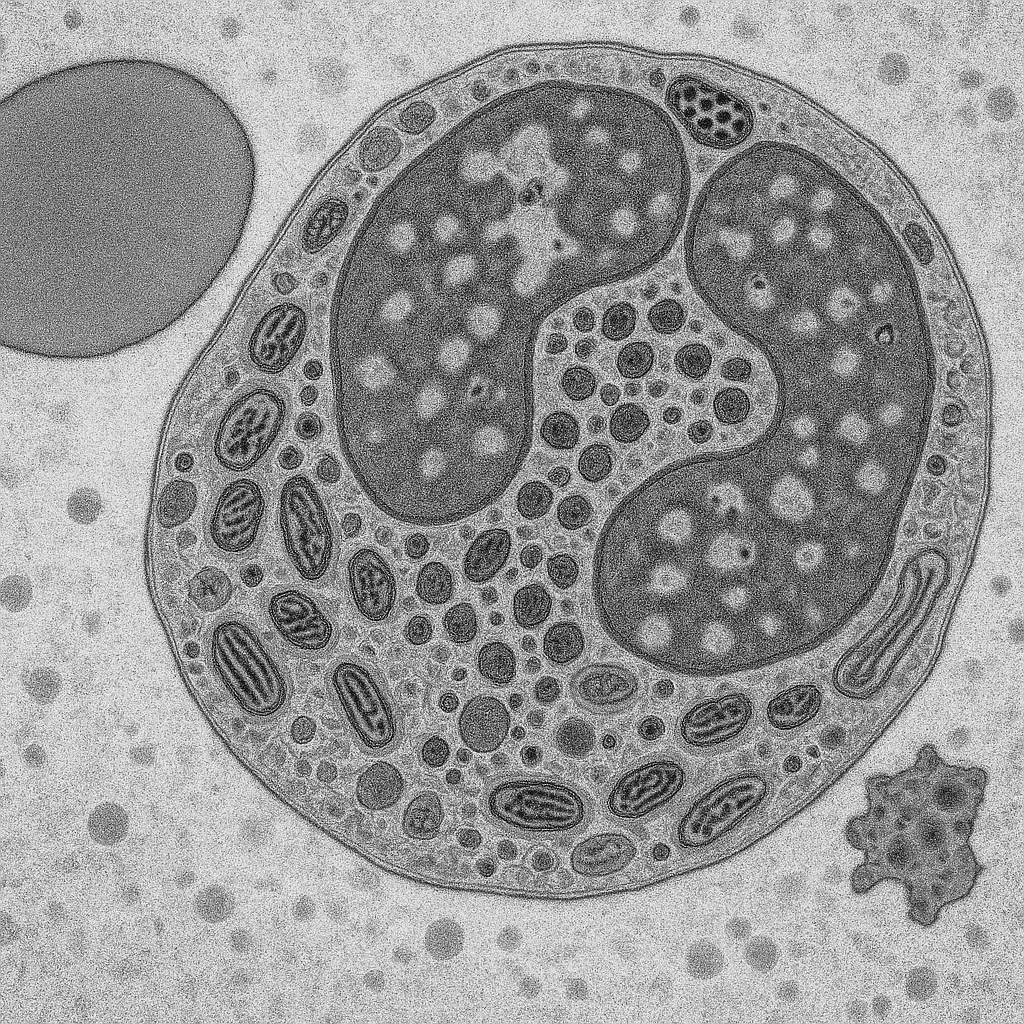
Formalin-fixed paraffin-embedded tissue section with hematoxylin and eosin staining
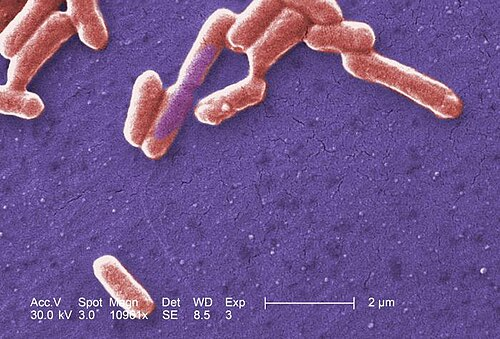
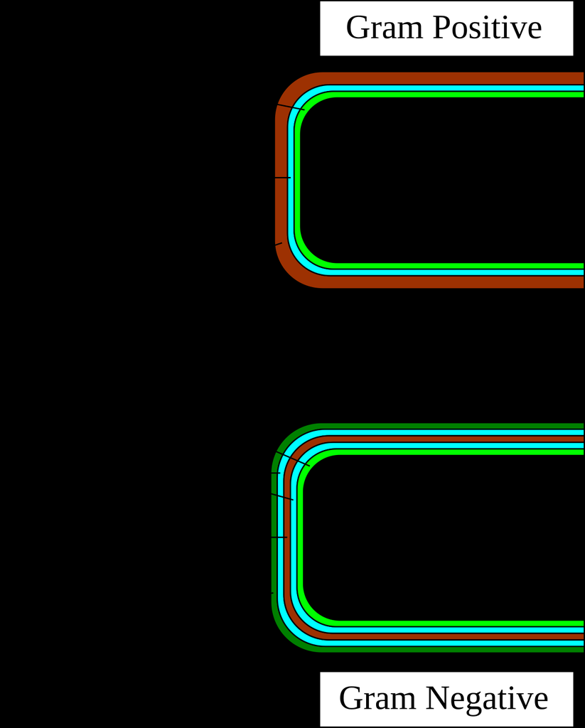
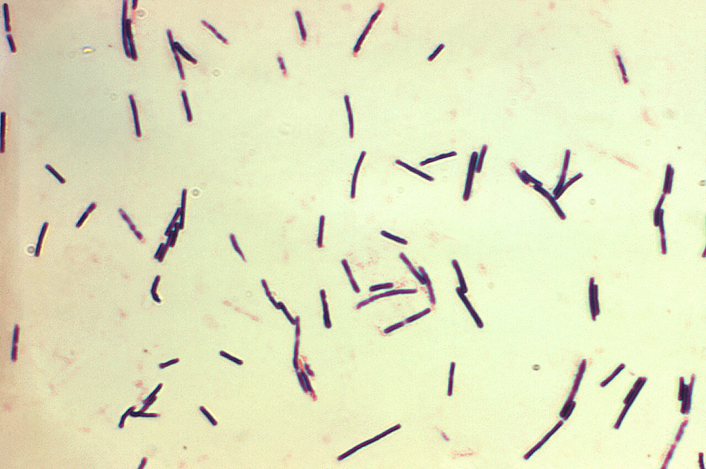
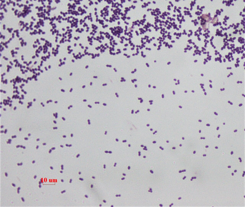
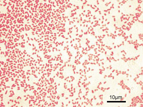
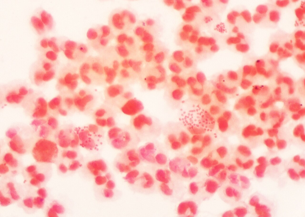
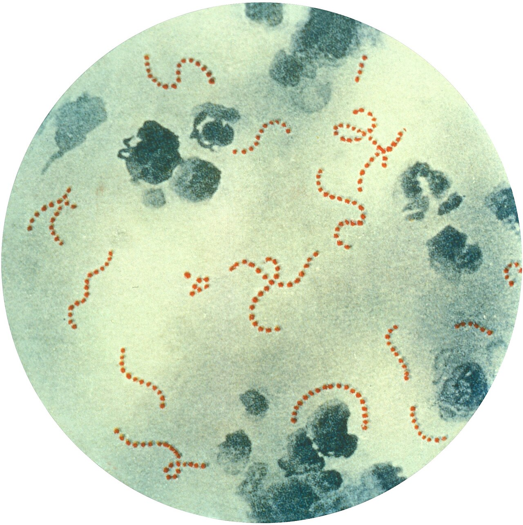
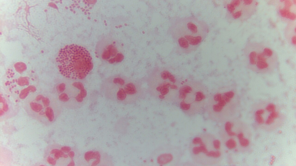
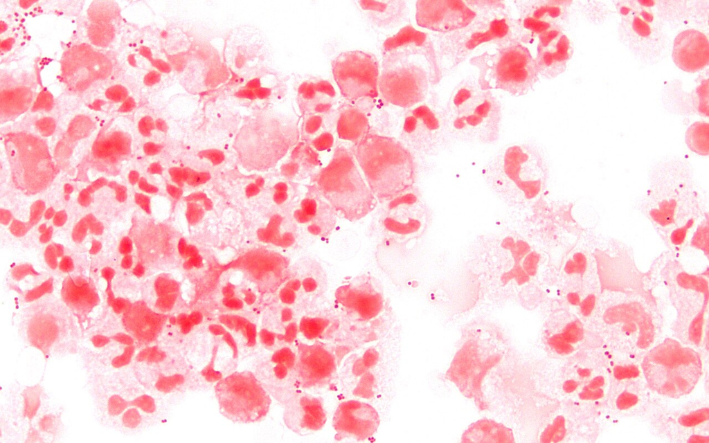
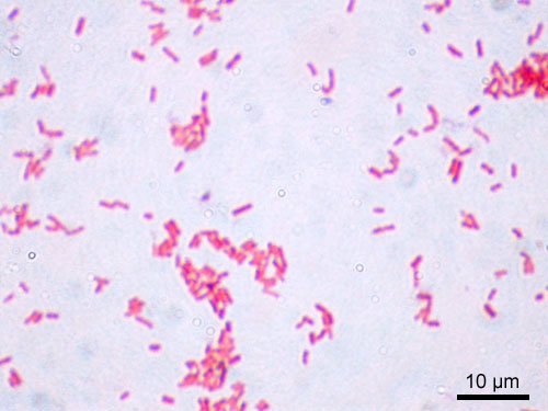
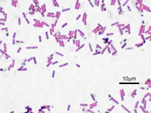
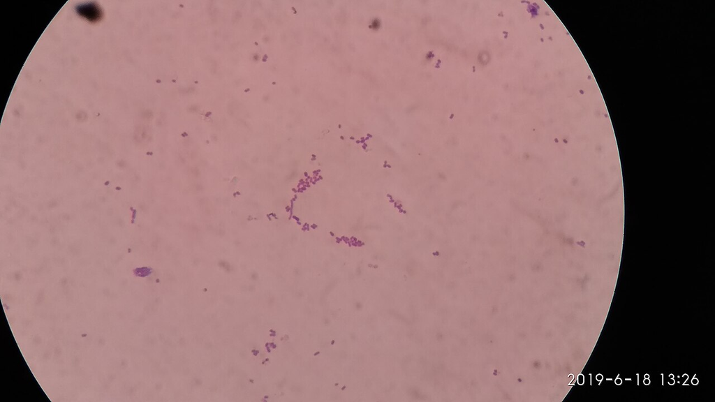
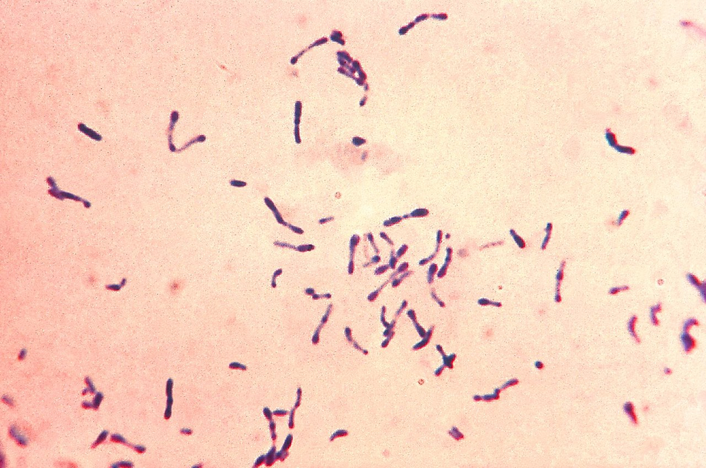
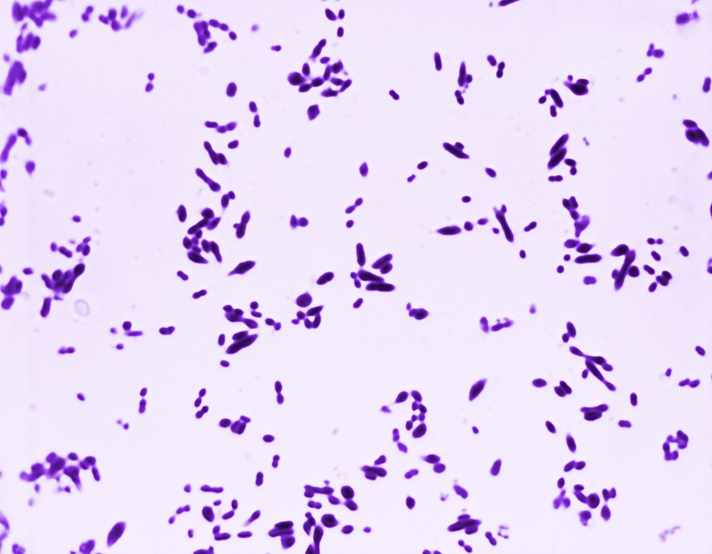
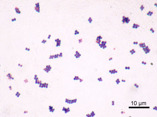
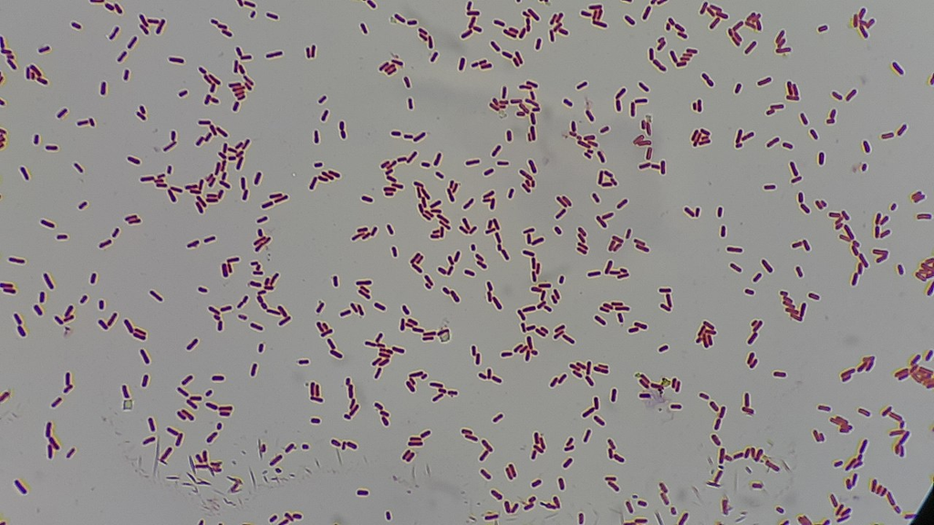
Heat-fixed bacterial smear with Gram staining showing gram-positive and gram-negative organisms

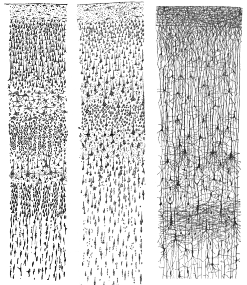

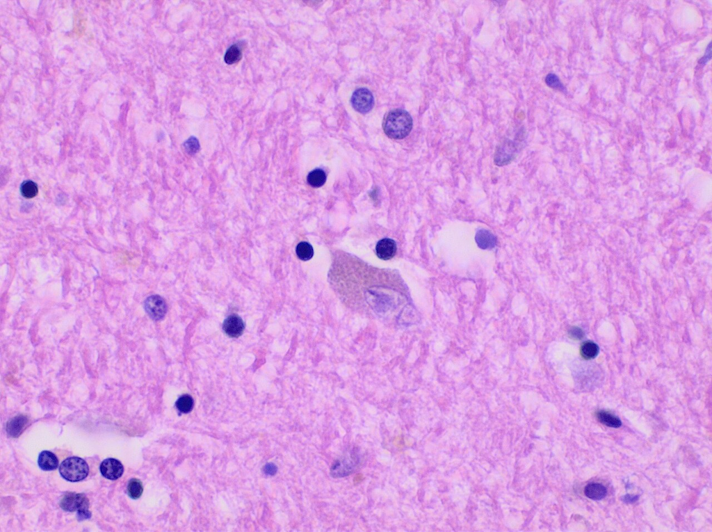
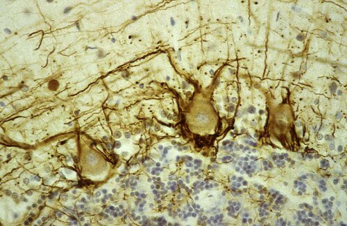
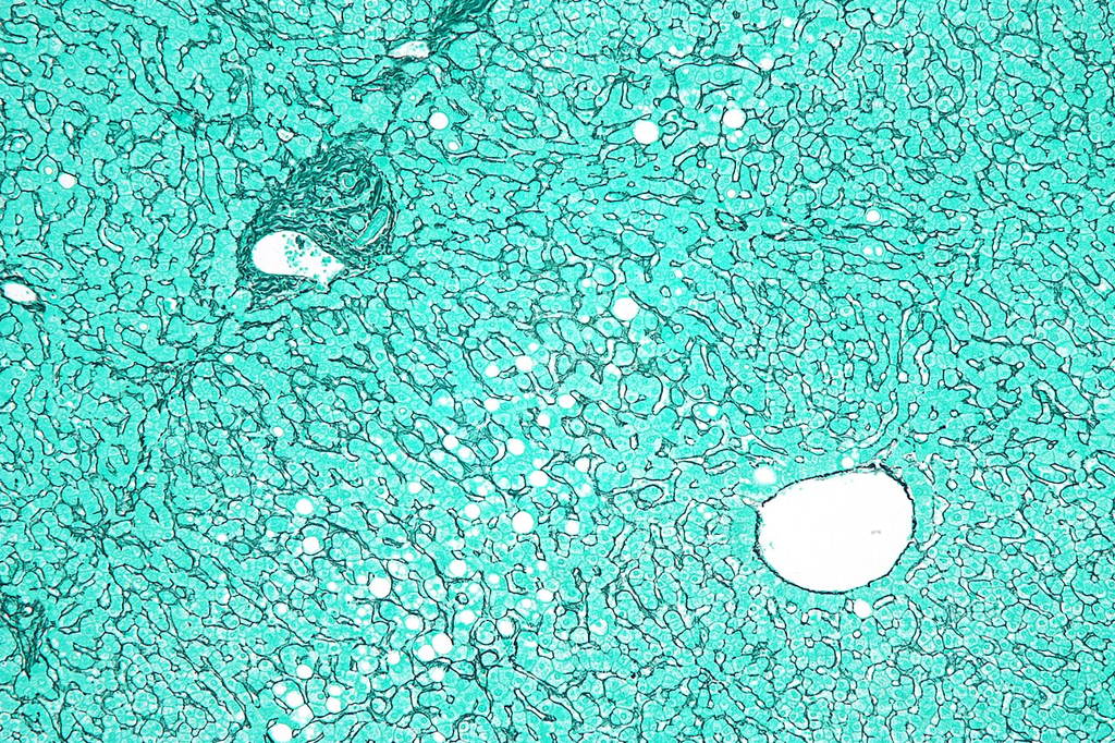
Golgi-stained brain tissue section showing neuronal morphology
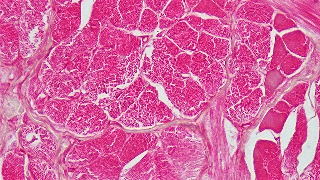
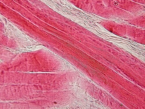
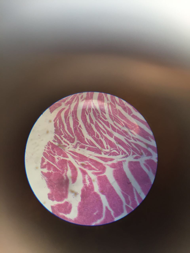
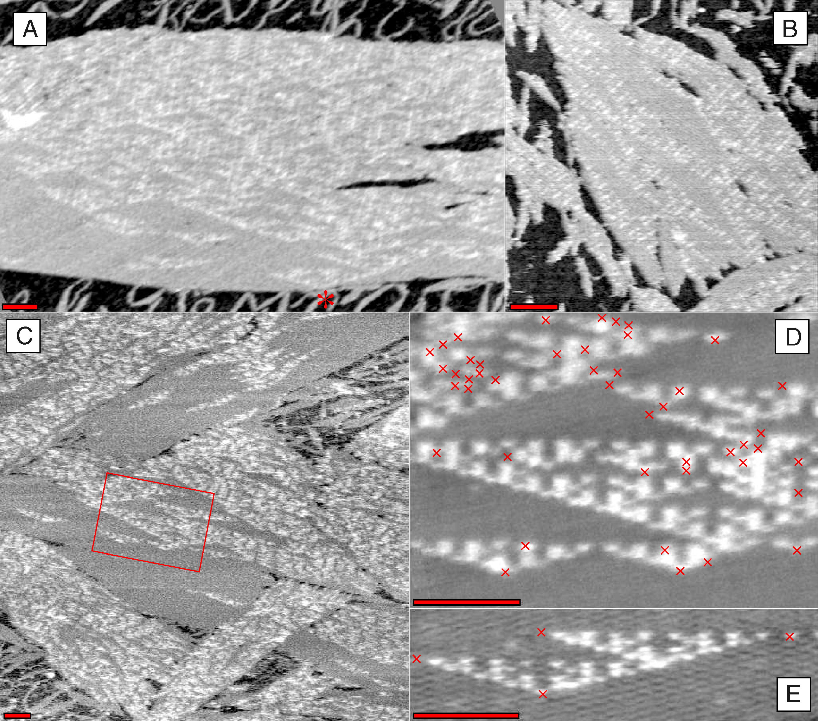
Transverse section of skeletal muscle showing fiber bundles and striations
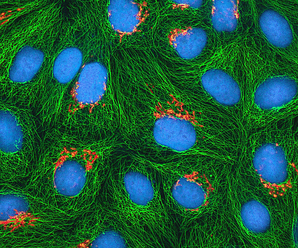
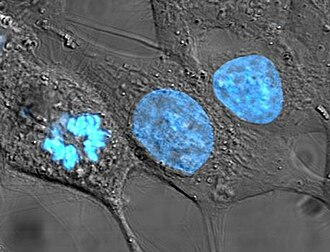
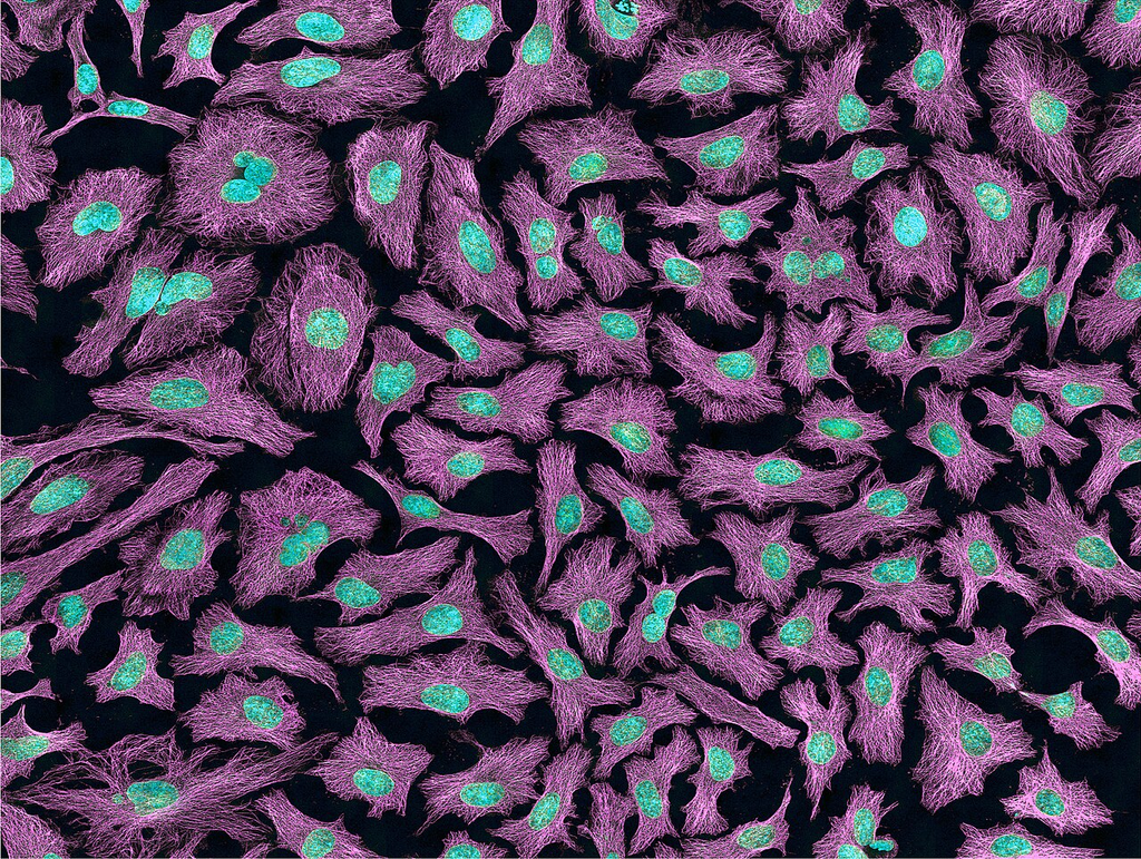
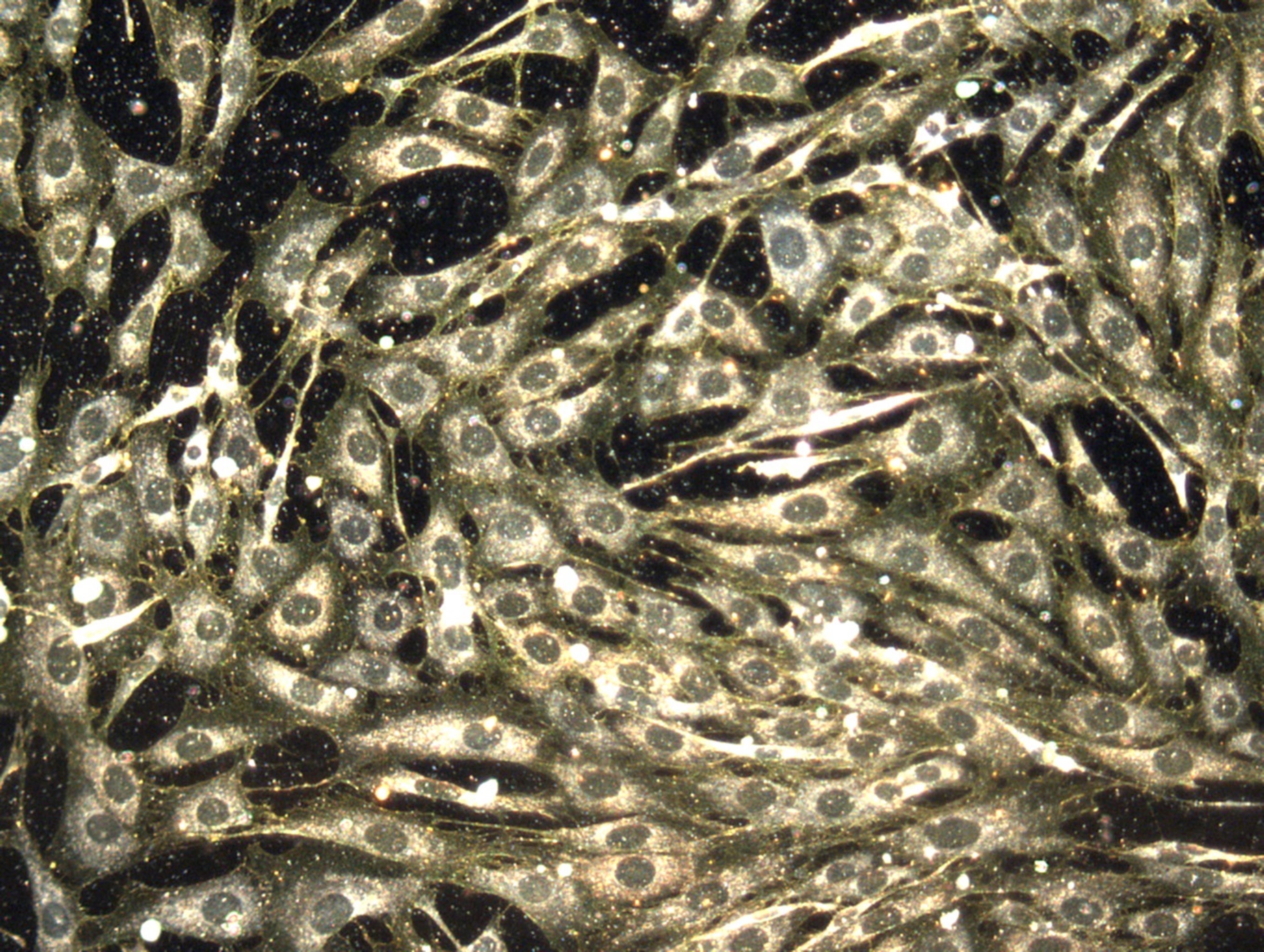
Cultured HeLa cells (cervical adenocarcinoma) in monolayer
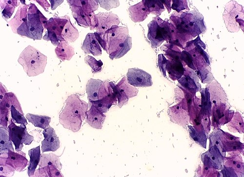
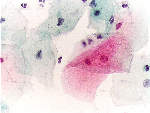
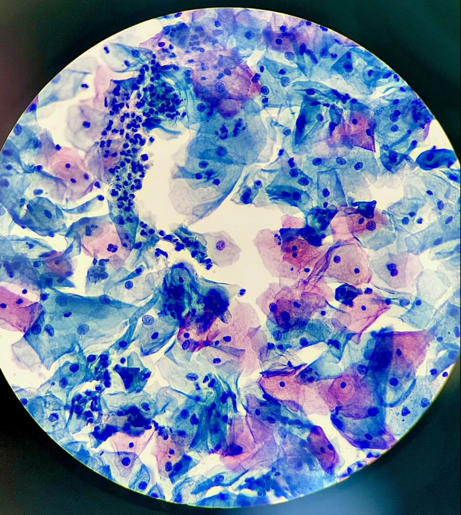
Papanicolaou-stained cervical smear for screening
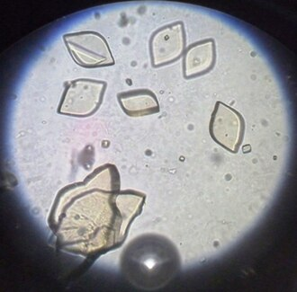
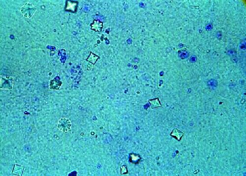
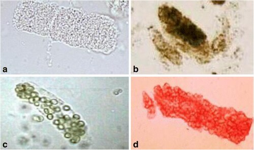
Centrifuged urine sediment showing casts, crystals, and cells
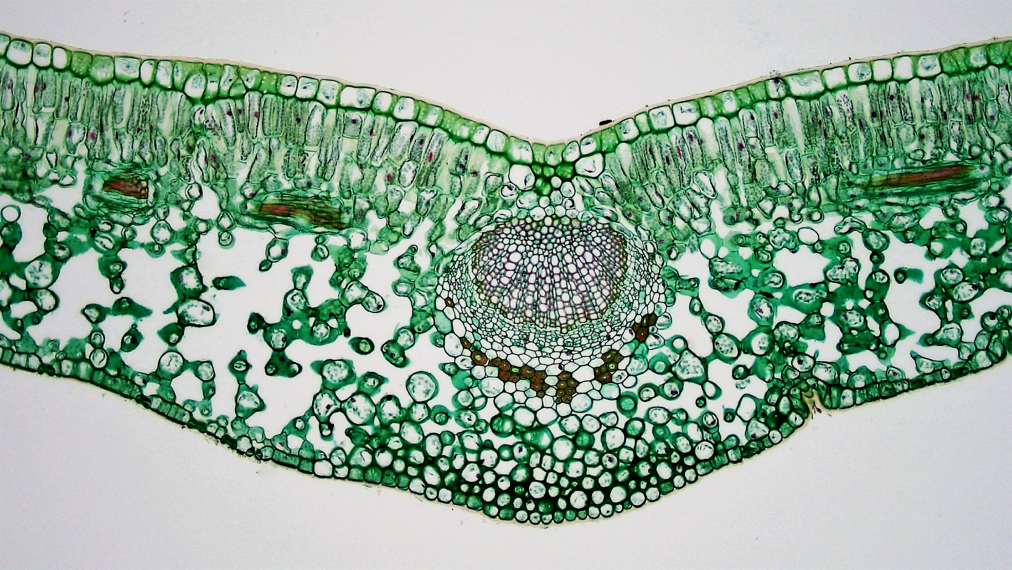
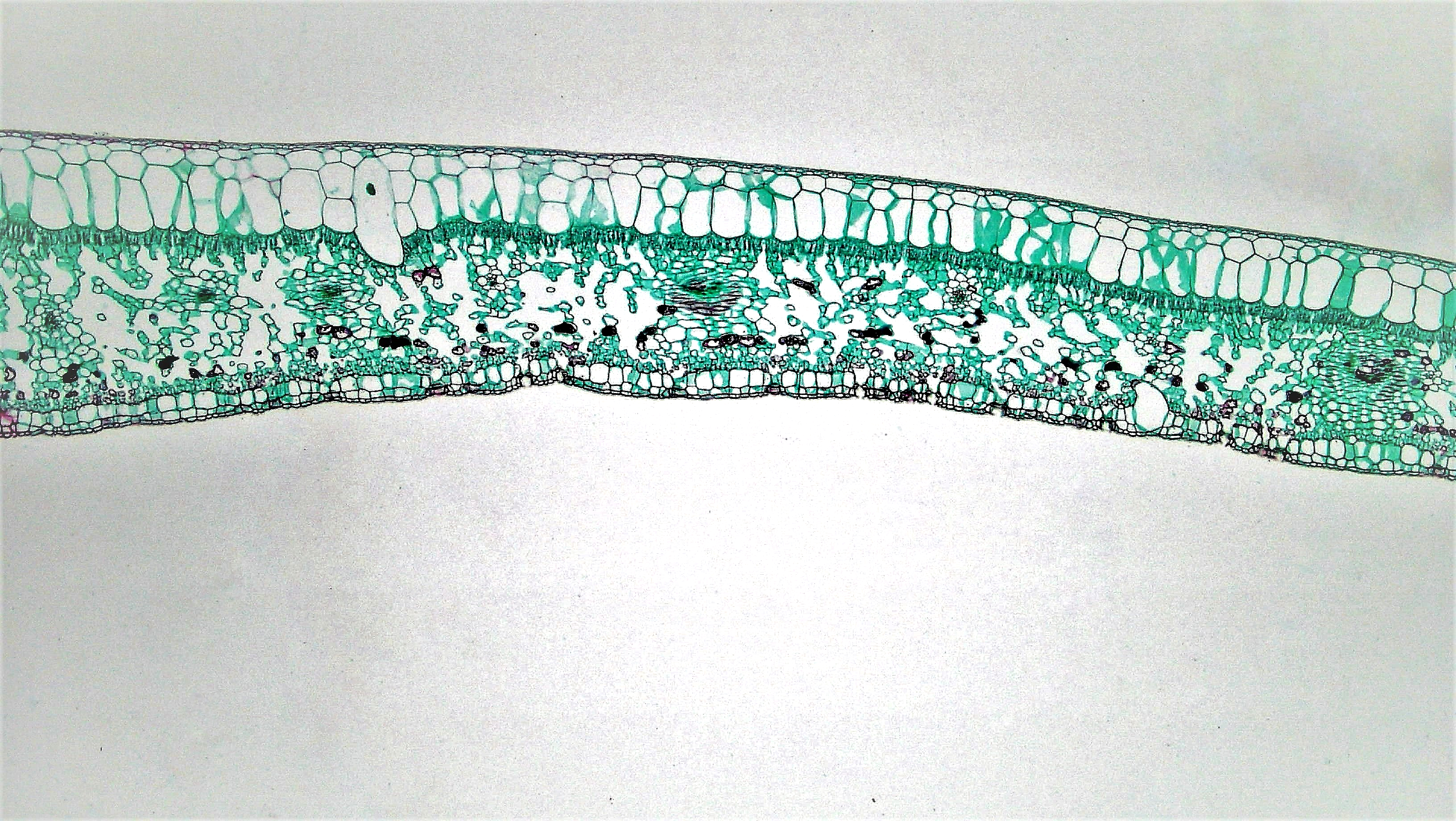
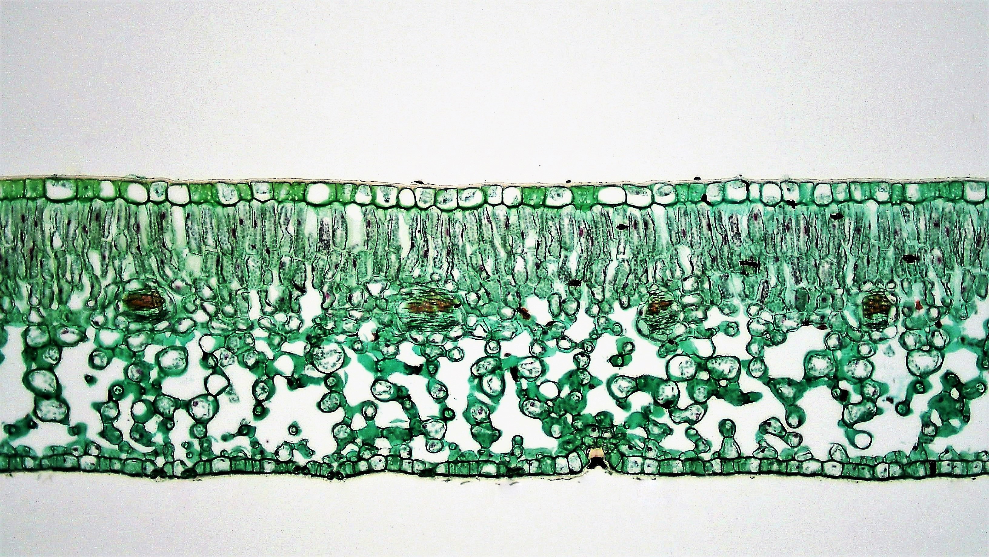
Transverse section of dicot leaf showing epidermis, mesophyll, and vascular bundles
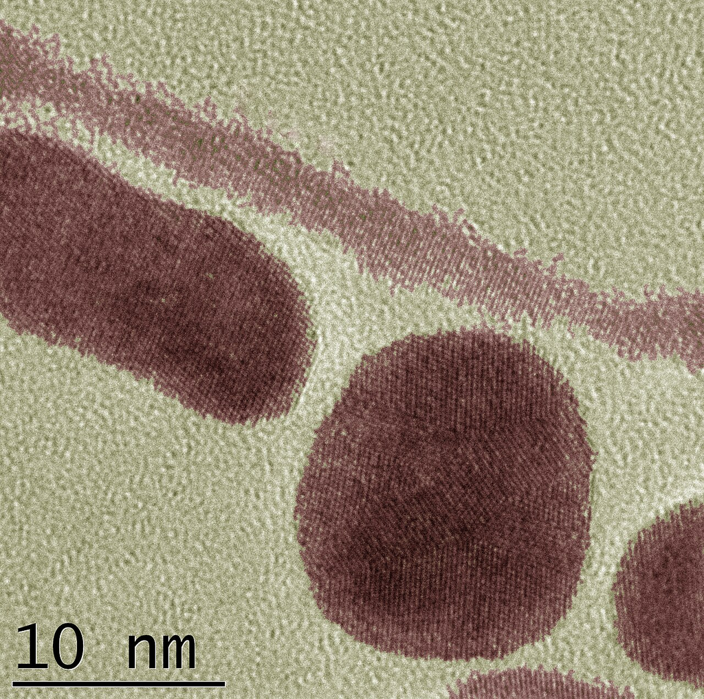
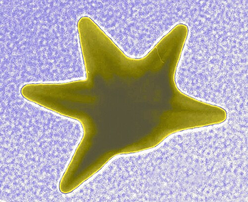
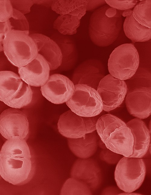
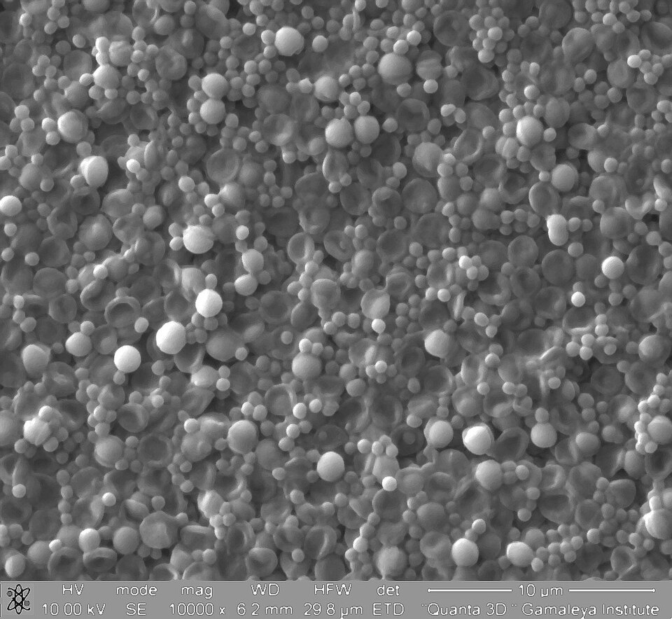
Gold nanoparticles (50 nm) on carbon grid for TEM imaging
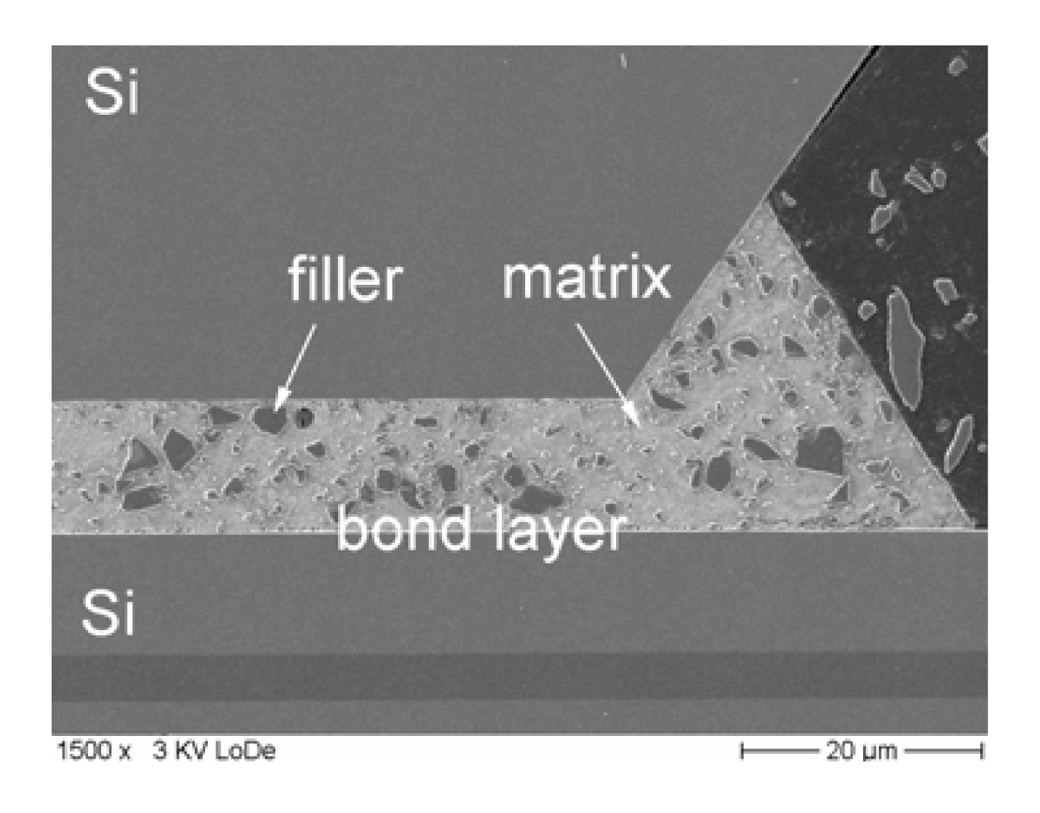
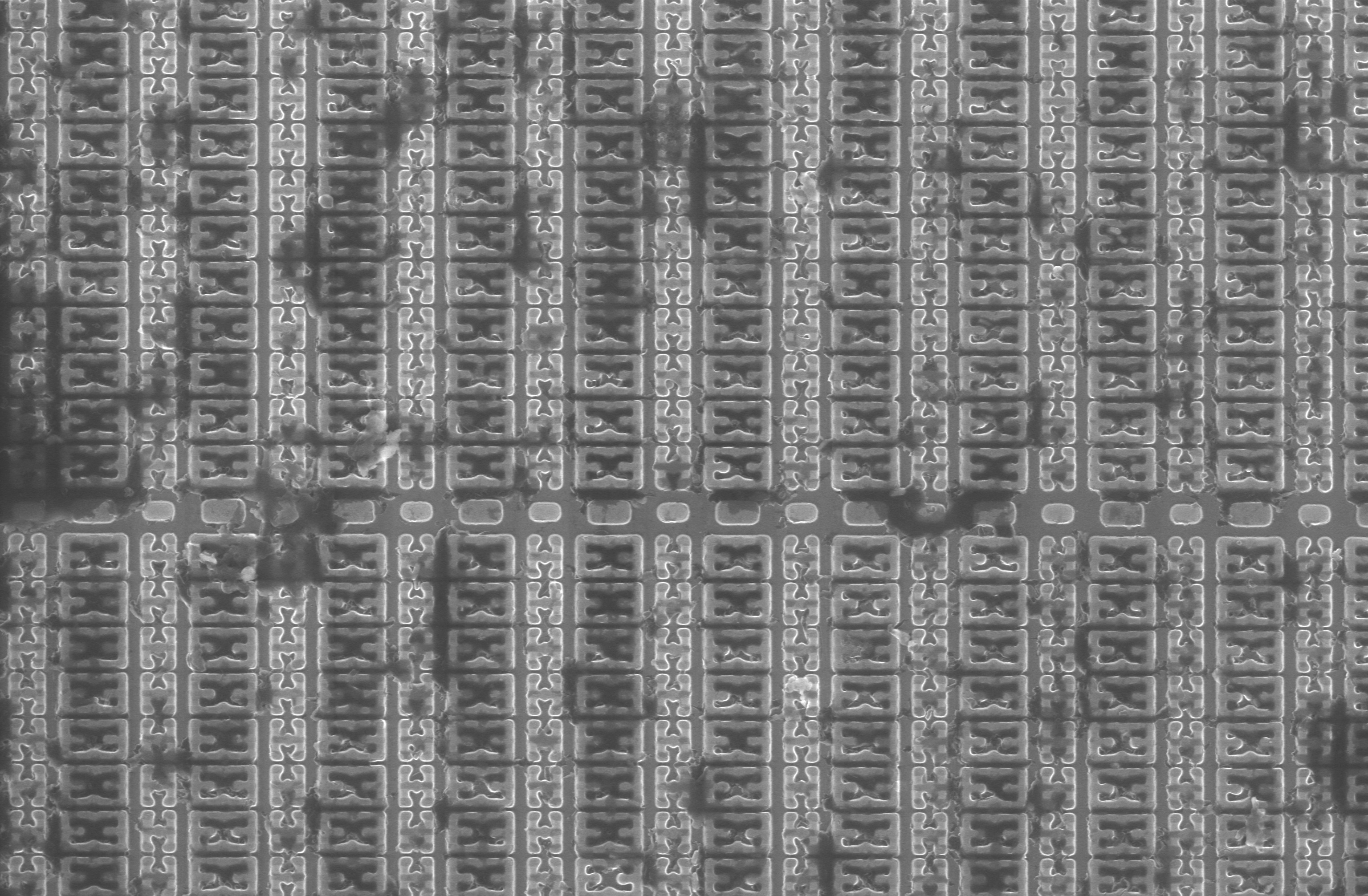
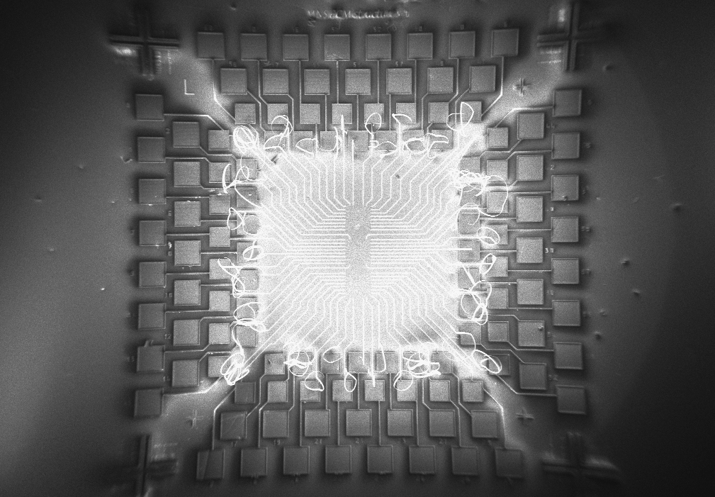
Silicon wafer cross-section showing transistor gate structures
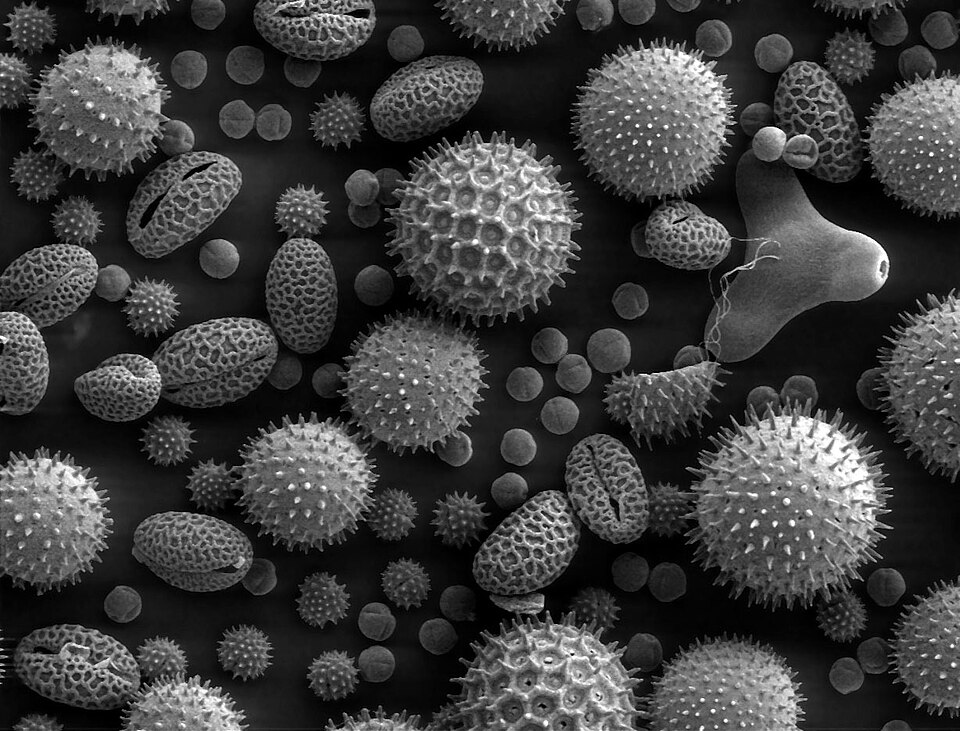
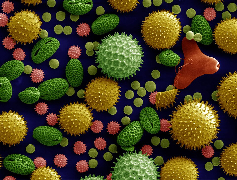
Acetolyzed mixed pollen from flowering plants
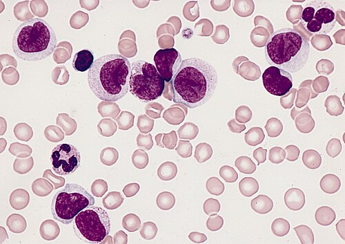

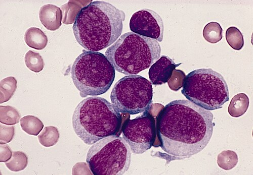
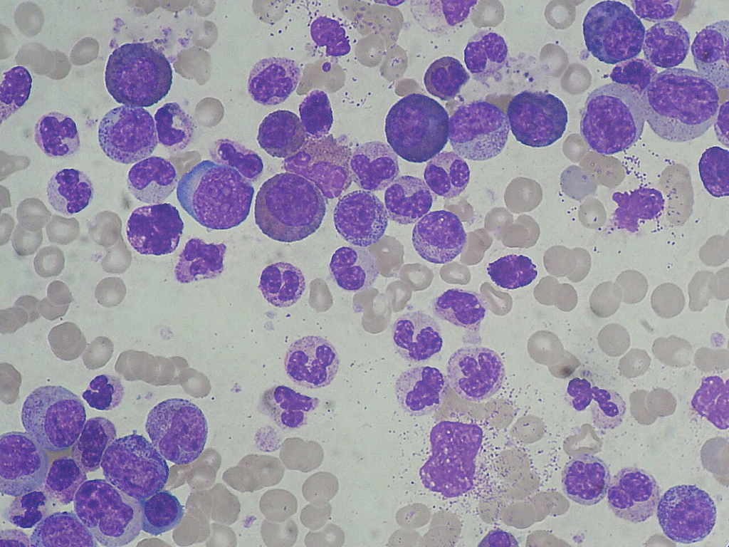
Wright-Giemsa stained bone marrow aspirate smear
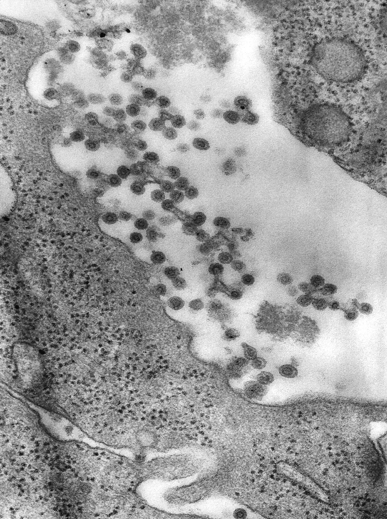
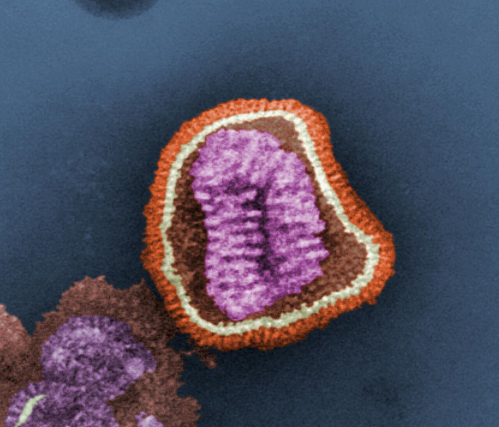
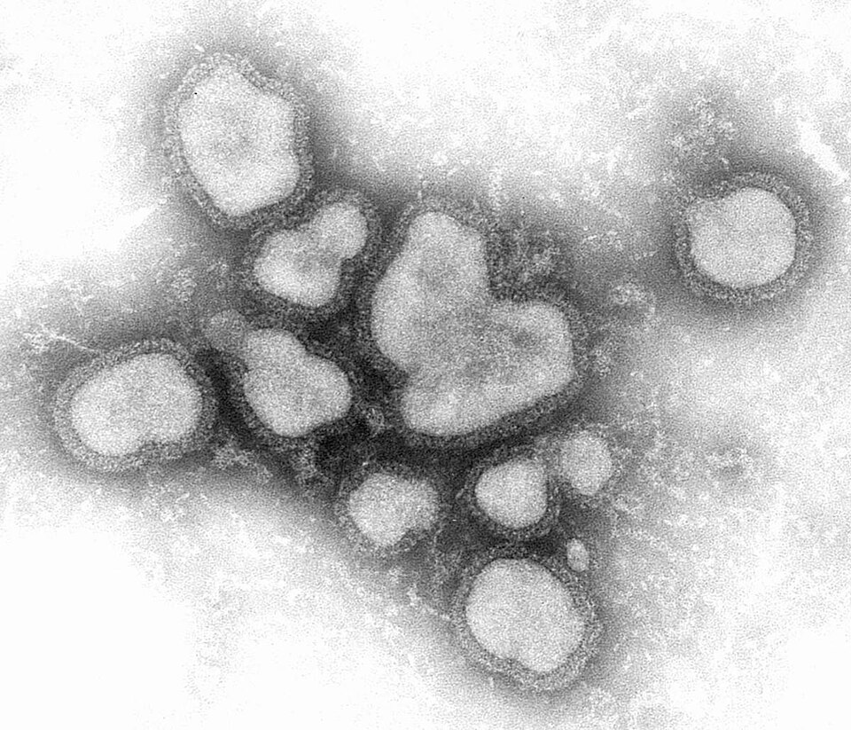
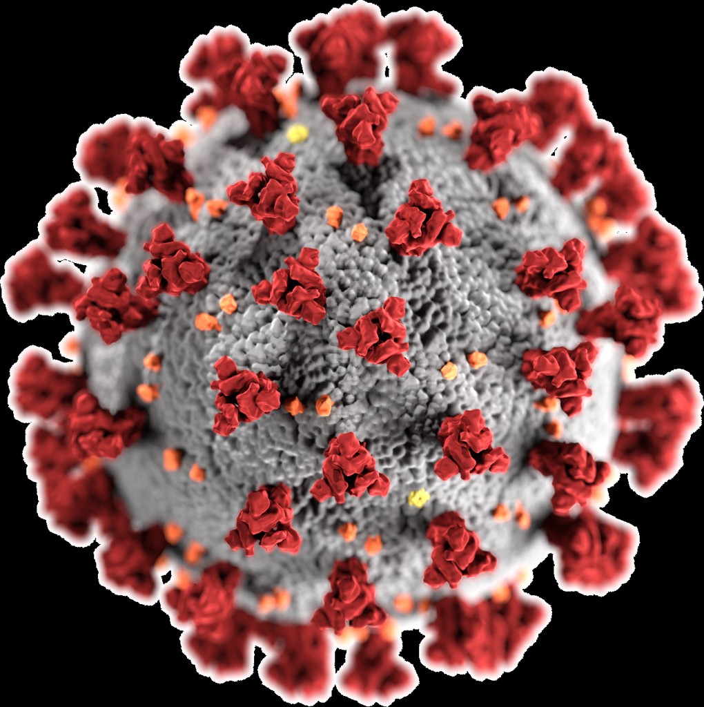
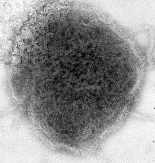
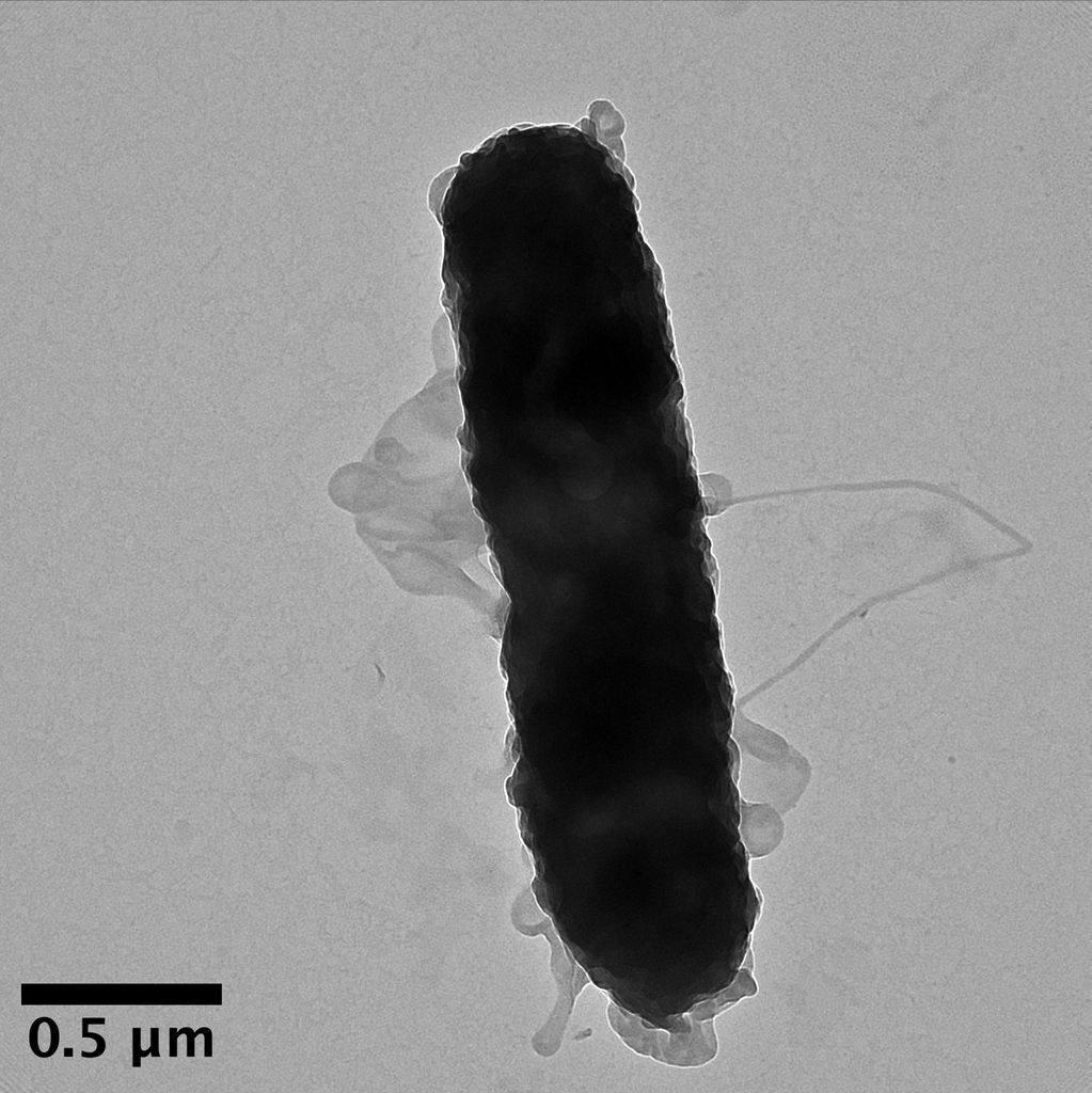
Negatively stained virus particles on carbon-coated copper grid
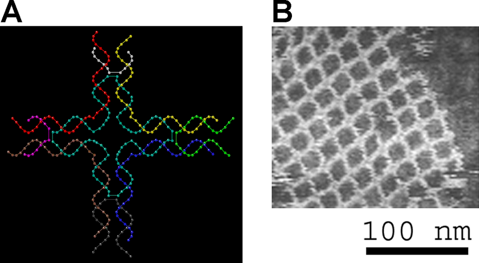
Designed DNA origami structures deposited on mica for AFM imaging
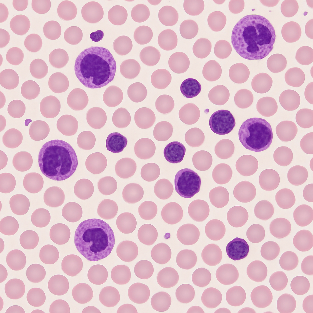
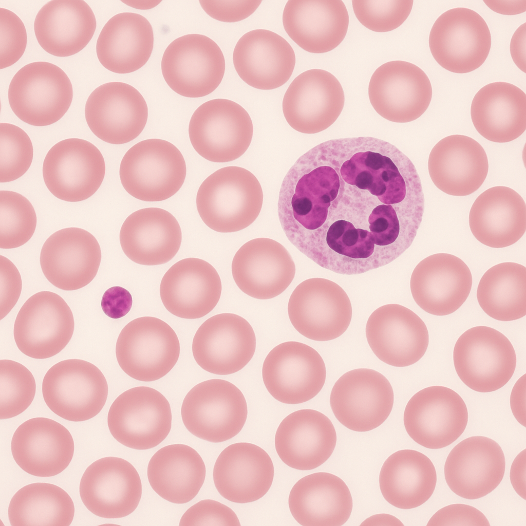
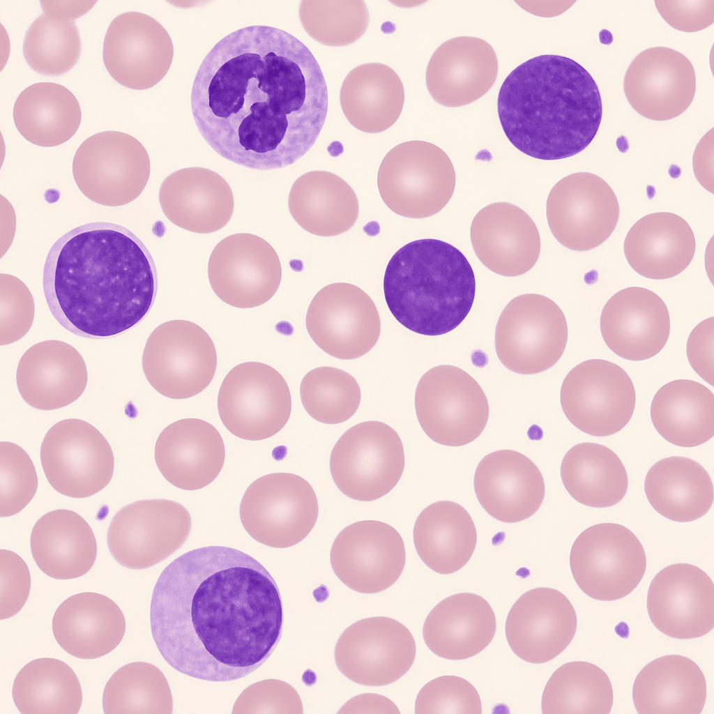
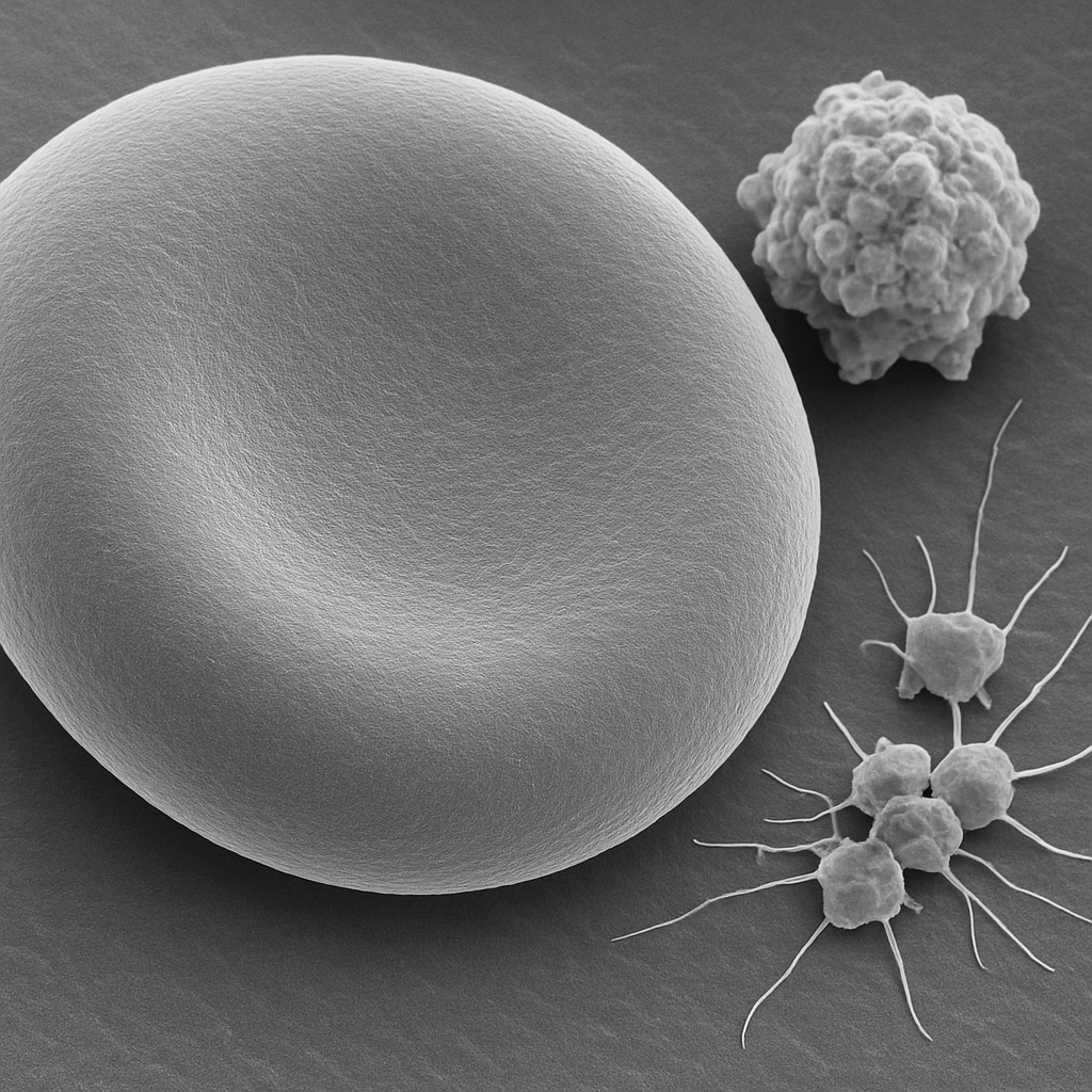
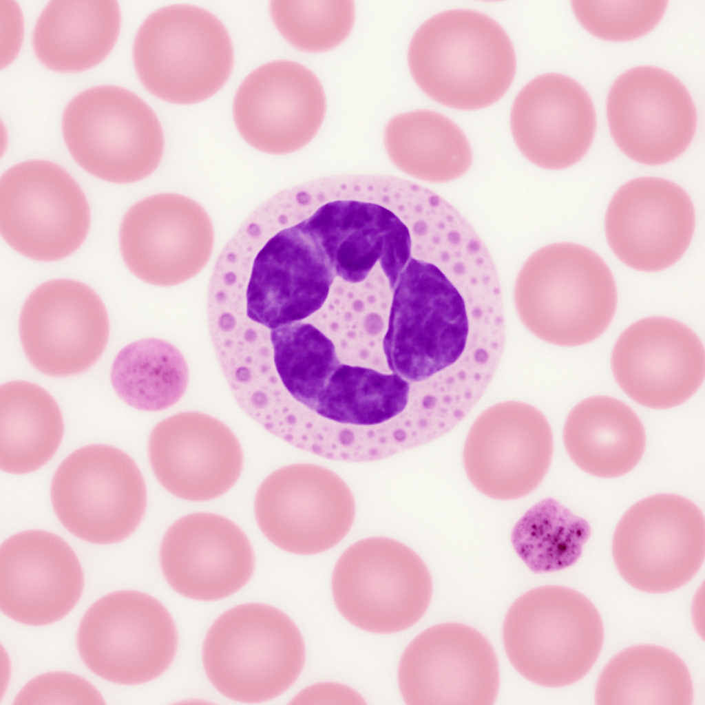

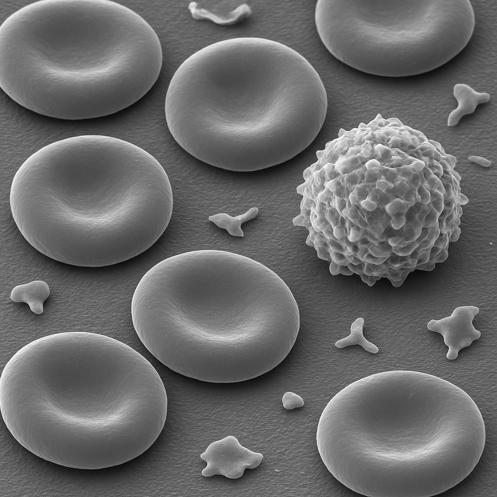
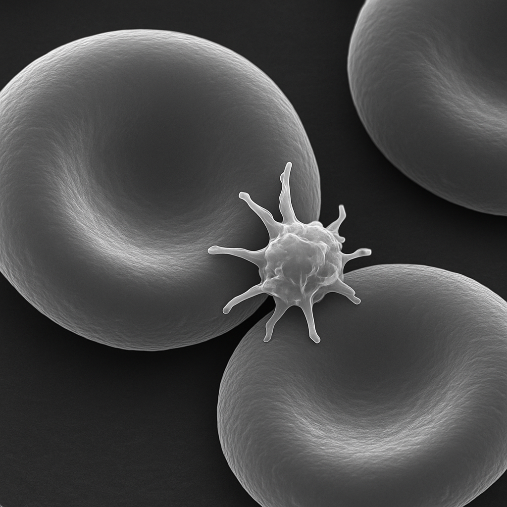
AI-generated peripheral blood smear image set spanning the full range of microscopy...
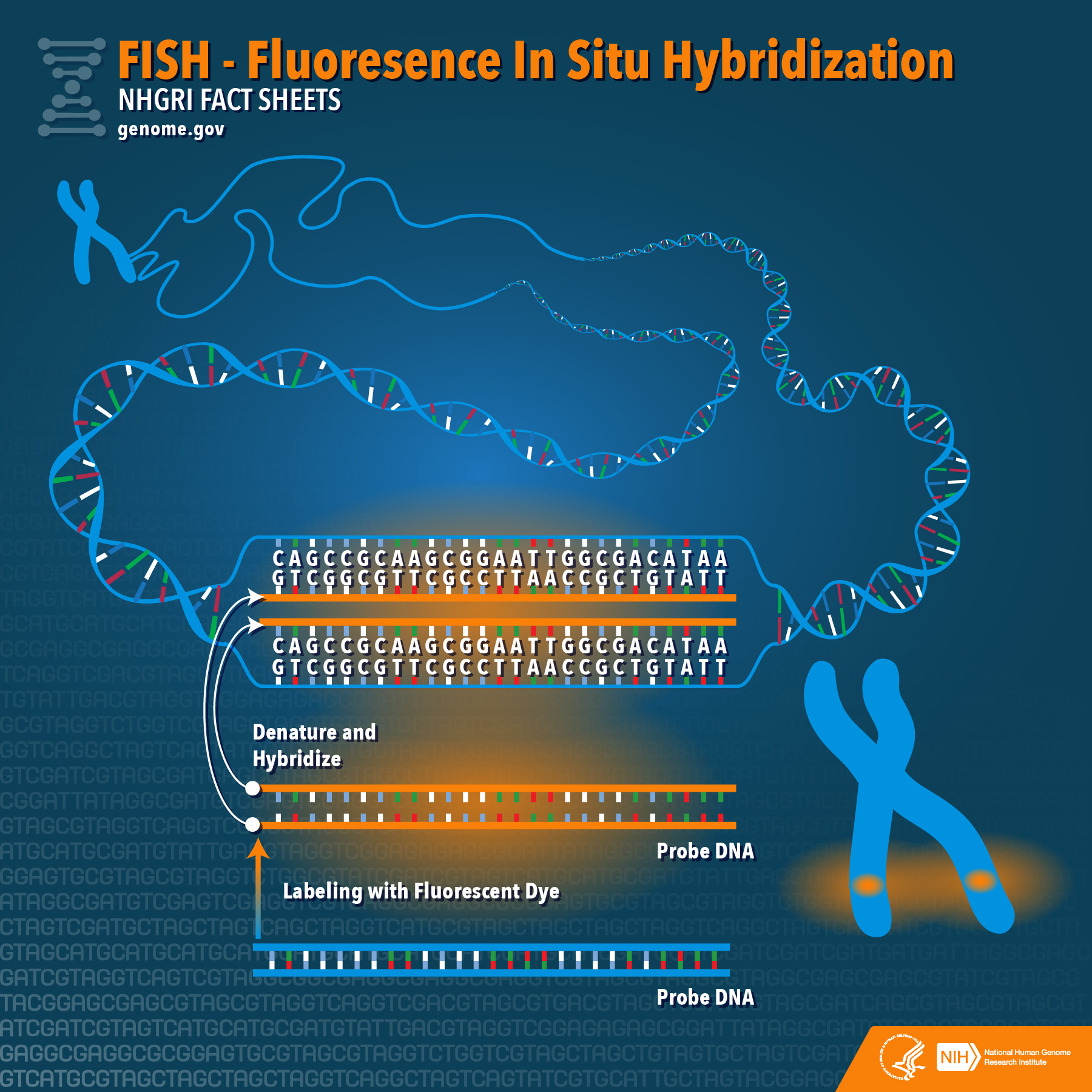
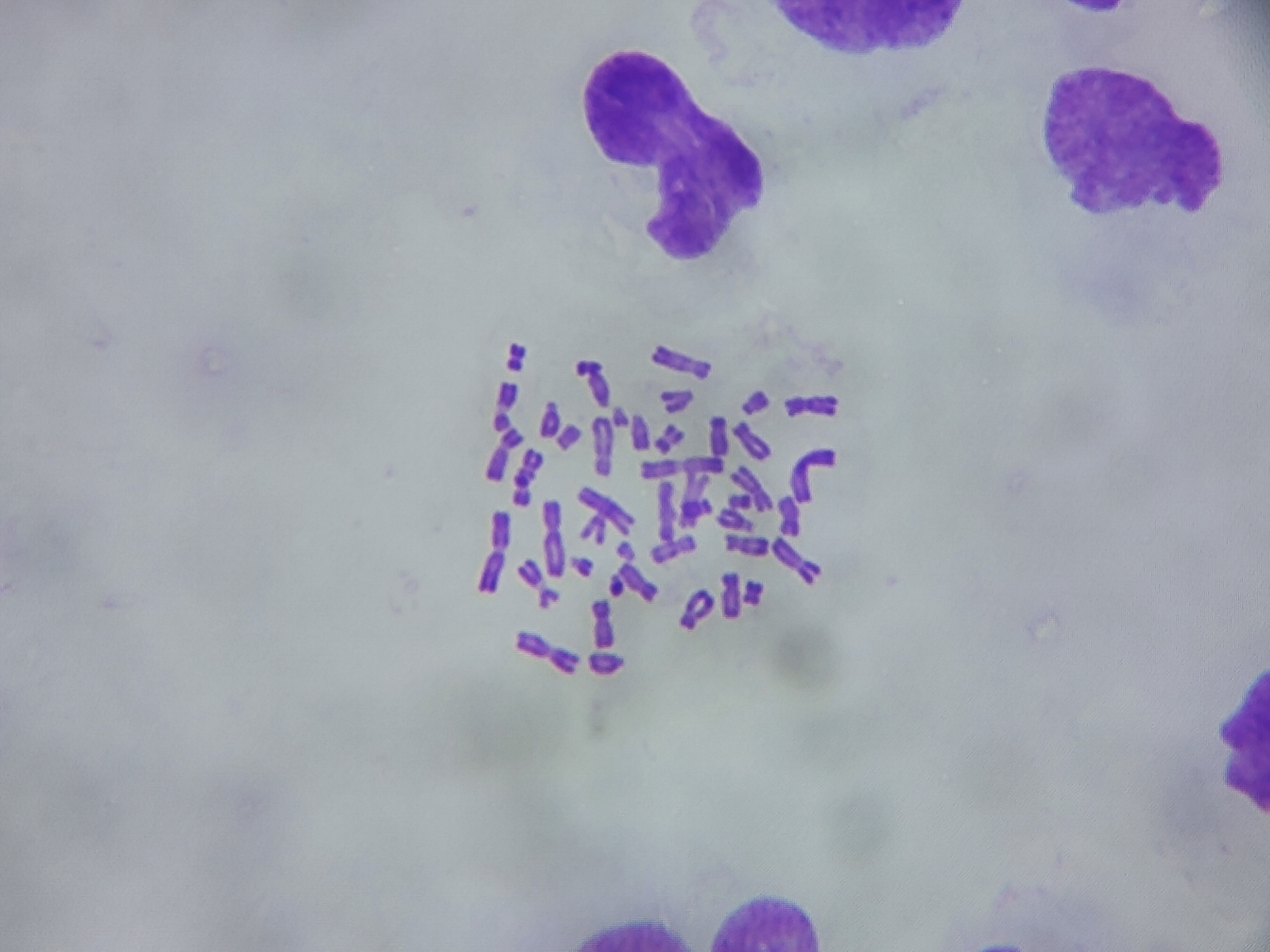
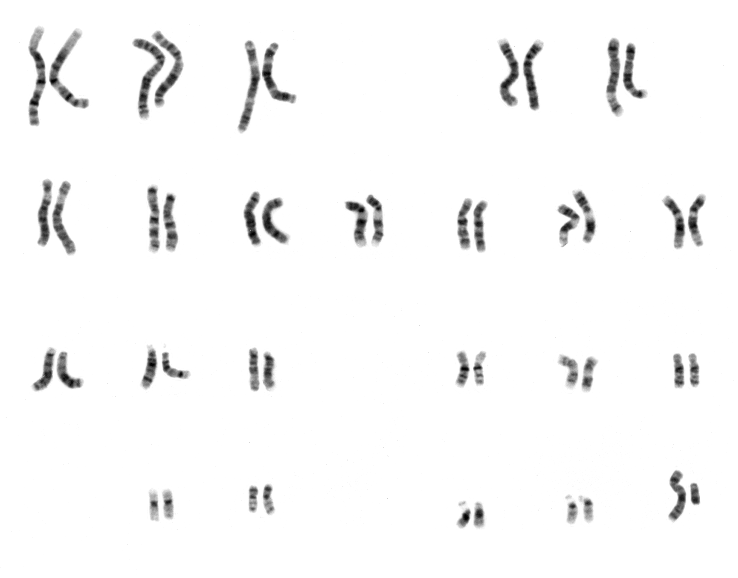
Human chromosome preparations for cytogenetic analysis including G-banded karyotypes,...
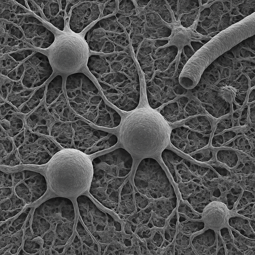
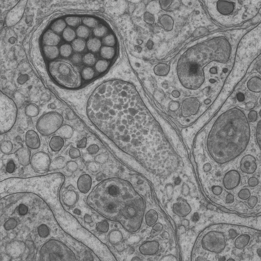
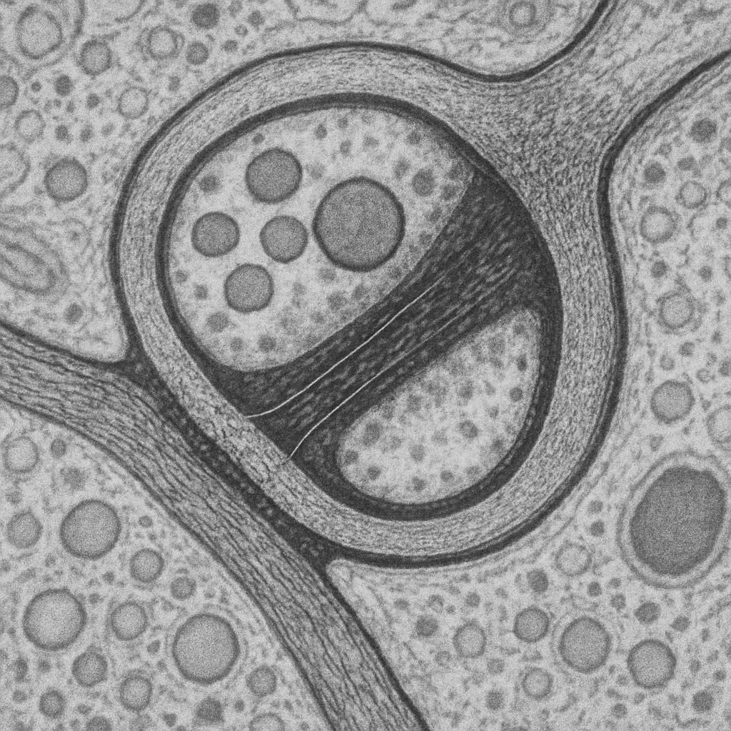

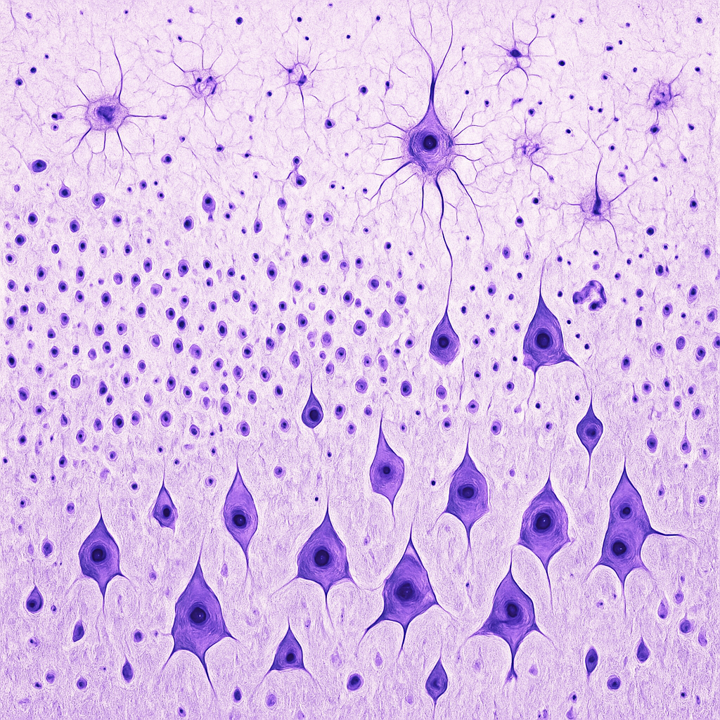
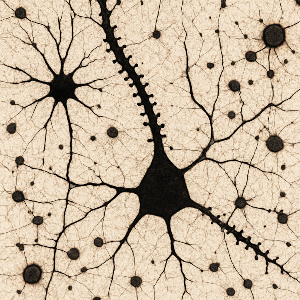
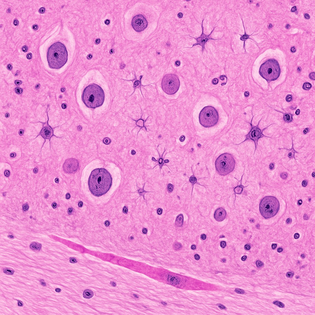
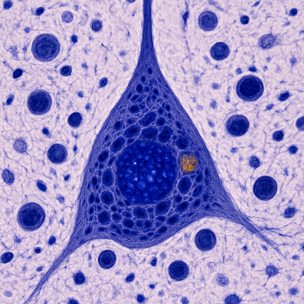
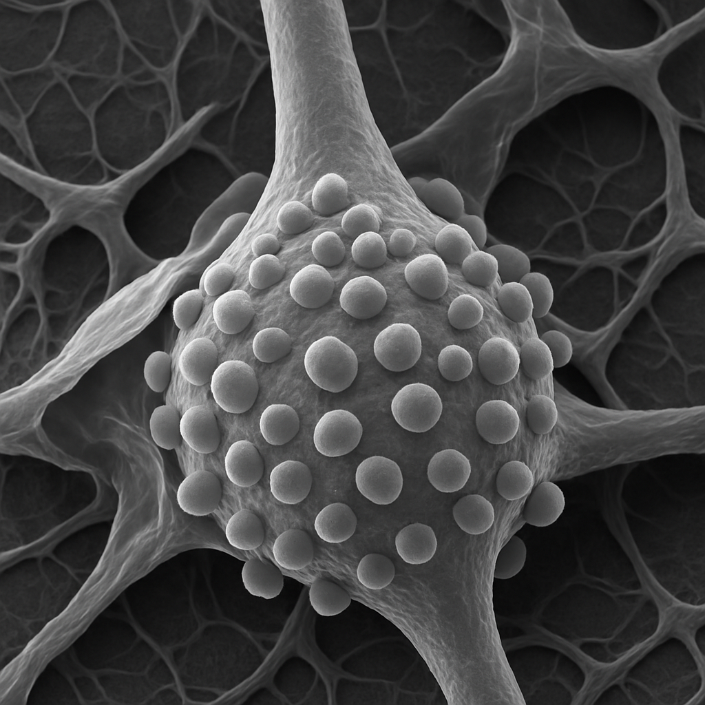
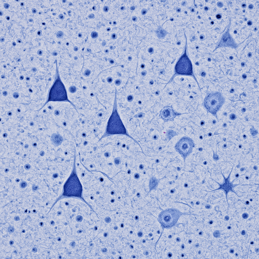
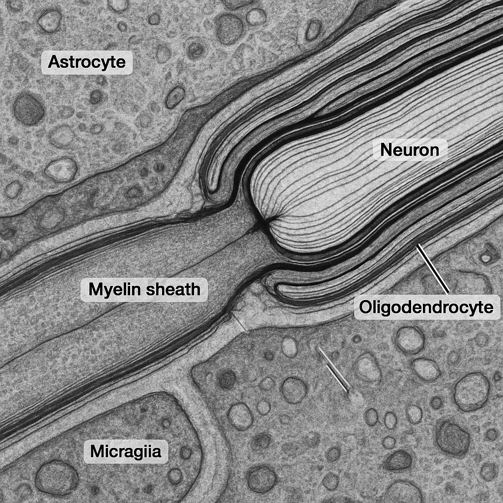
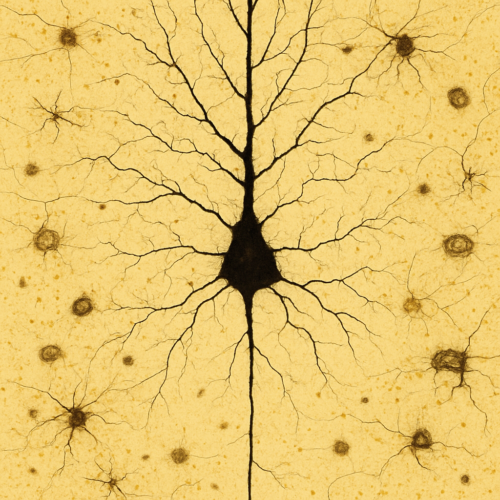
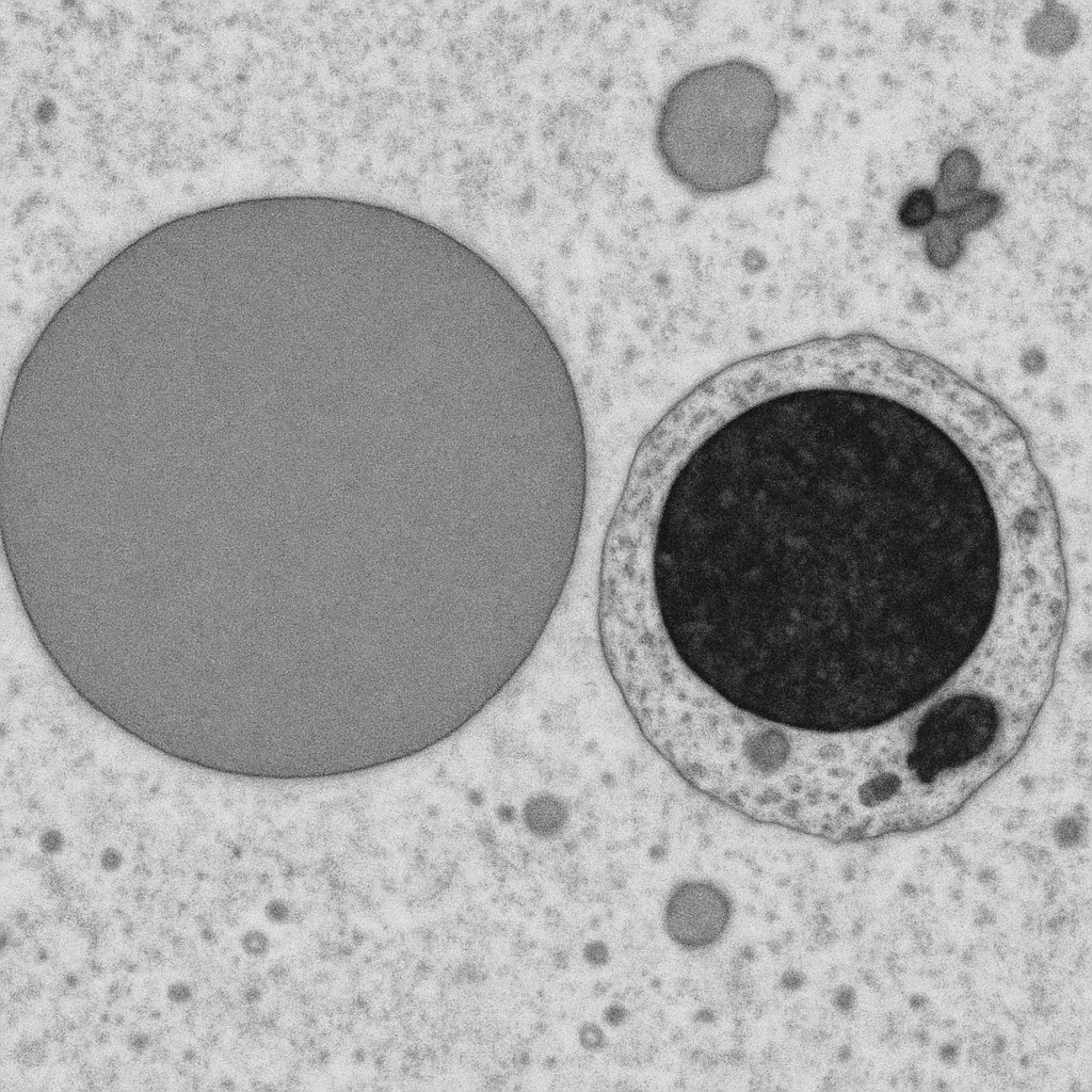
AI-generated brain tissue image set spanning the full range of microscopy...