Skeletal Muscle Cross-Section
Skeletal Muscle Cross-Section
HistologyTransverse section of skeletal muscle showing fiber bundles and striations
Drag & drop images here to add to this specimen
or click Upload Image aboveRelease to upload
Image Library
3 images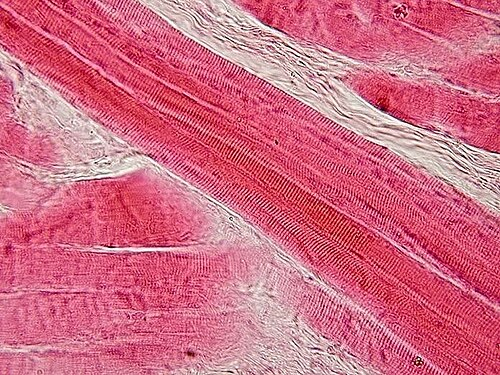
Longitudinal section of skeletal muscle tissue stained with H&E showing parallel muscle fibers with visible cross-striations. Peripheral nuclei are flattened against the sarcolemma. The banding pattern (A-bands and I-bands) is visible at higher magnification.
H&E stained — longitudinal fibers showing striations
Wikimedia Commons — CC BY-SA 3.0
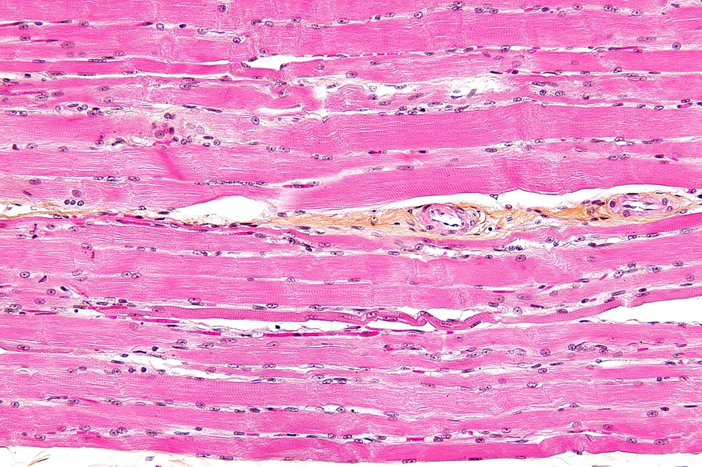
High-magnification view of skeletal muscle fibers in H&E-stained section showing distinct cross-striations, multinucleated fibers, and connective tissue endomysium between individual muscle cells.
H&E stained — high-mag showing striations and peripheral nuclei
Wikimedia Commons — CC BY-SA 3.0
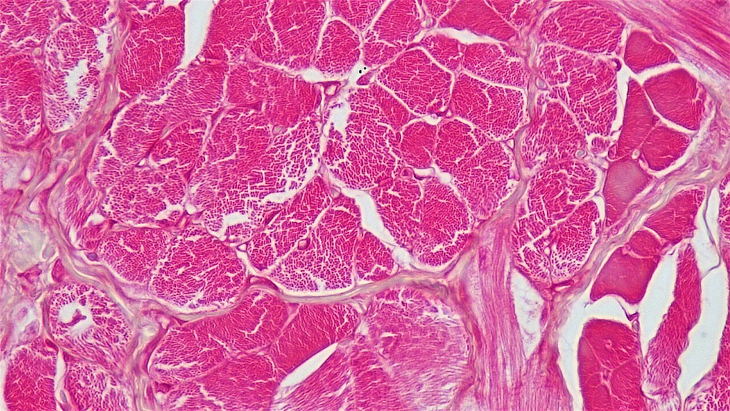
Transverse section of skeletal muscle stained with H&E showing polygonal muscle fibers in cross-section. Each fiber shows peripheral nuclei and endomysium separating individual fibers. Fascicles are bounded by perimysium.
H&E stained — classic skeletal muscle cross-section morphology
Wikimedia Commons — CC BY-SA 3.0
Compatible Microscopes
| Model | Manufacturer | Type | Magnification Range | NA Max | Resolution |
|---|---|---|---|---|---|
| DM6 B | Leica Microsystems | Upright Optical | 1.25–100× | 1.4 | 200 nm |
| DMi8 | Leica Microsystems | Inverted Optical | 2.5–100× | 1.47 | 185 nm |
| Eclipse Ti2 | Nikon | Inverted Optical | 2–100× | 1.45 | 190 nm |
| Eclipse Ni | Nikon | Upright Optical | 2–100× | 1.4 | 200 nm |
| BX53 | Olympus (Evident) | Upright Optical | 2–100× | 1.4 | 200 nm |
| IX83 | Olympus (Evident) | Inverted Optical | 2–100× | 1.4 | 200 nm |
| Axio Observer 7 | Zeiss | Inverted Optical | 5–100× | 1.4 | 200 nm |
| Axio Imager 2 | Zeiss | Upright Optical | 1.25–100× | 1.4 | 200 nm |
| Primostar 3 | Zeiss | Upright Optical | 4–100× | 1.25 | 350 nm |
Edit Image Metadata
AI Generate Image — Skeletal Muscle Cross-Section
Fill in the imaging criteria below. A detailed prompt will be built from your selections and sent to OpenAI to generate a realistic microscopy image.