Pap Smear (Cervical Cytology)
Pap Smear (Cervical Cytology)
CytologyPapanicolaou-stained cervical smear for screening
Drag & drop images here to add to this specimen
or click Upload Image aboveRelease to upload
Image Library
3 images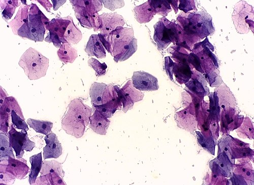
Normal Papanicolaou-stained cervical smear showing superficial and intermediate squamous epithelial cells. Cells are flat with small pyknotic nuclei and abundant pale cytoplasm. No dysplastic changes.
Normal cytology — NILM (Negative for Intraepithelial Lesion or Malignancy)
Wikimedia Commons — Public Domain
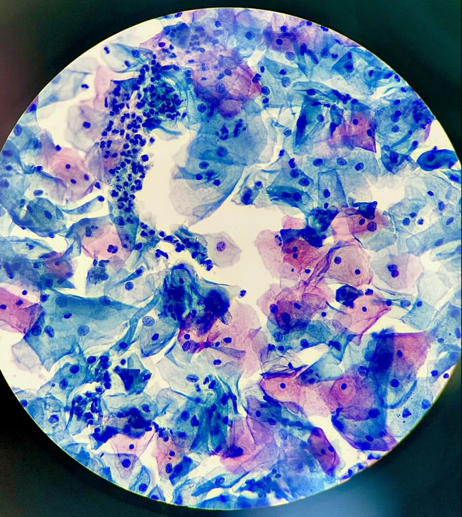
Papanicolaou-stained cervical cytology smear showing epithelial cells with characteristic staining pattern. Cytoplasm stains in shades of green and pink while nuclei appear deep blue-purple.
Pap stain — standard cervical screening cytology preparation
Wikimedia Commons — CC BY-SA 3.0
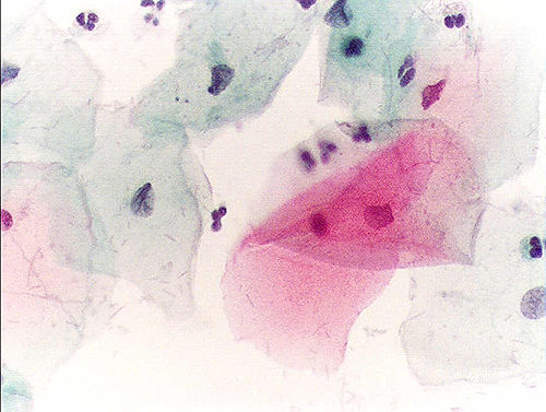
Normal Pap test showing mature squamous epithelial cells with abundant cytoplasm and small regular nuclei. No atypical cells or dysplastic changes present. Consistent with NILM classification.
Normal squamous cells — small nuclei, abundant cytoplasm, no atypia
Wikimedia Commons — CC BY-SA 3.0
Compatible Microscopes
| Model | Manufacturer | Type | Magnification Range | NA Max | Resolution |
|---|---|---|---|---|---|
| VHX-7000 | Keyence | Digital Microscope | 0.1–6000× | — | 100 nm |
| DM6 B | Leica Microsystems | Upright Optical | 1.25–100× | 1.4 | 200 nm |
| Eclipse Ni | Nikon | Upright Optical | 2–100× | 1.4 | 200 nm |
| BX53 | Olympus (Evident) | Upright Optical | 2–100× | 1.4 | 200 nm |
| Axio Imager 2 | Zeiss | Upright Optical | 1.25–100× | 1.4 | 200 nm |
| Primostar 3 | Zeiss | Upright Optical | 4–100× | 1.25 | 350 nm |
Edit Image Metadata
AI Generate Image — Pap Smear (Cervical Cytology)
Fill in the imaging criteria below. A detailed prompt will be built from your selections and sent to OpenAI to generate a realistic microscopy image.