Plant Leaf Cross-Section
Plant Leaf Cross-Section
BotanyTransverse section of dicot leaf showing epidermis, mesophyll, and vascular bundles
Drag & drop images here to add to this specimen
or click Upload Image aboveRelease to upload
Image Library
3 images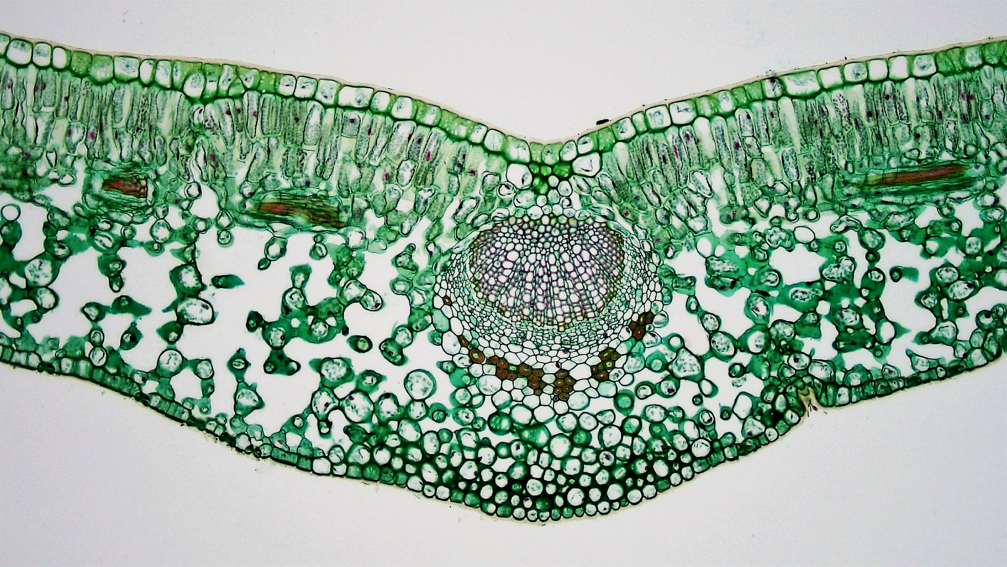
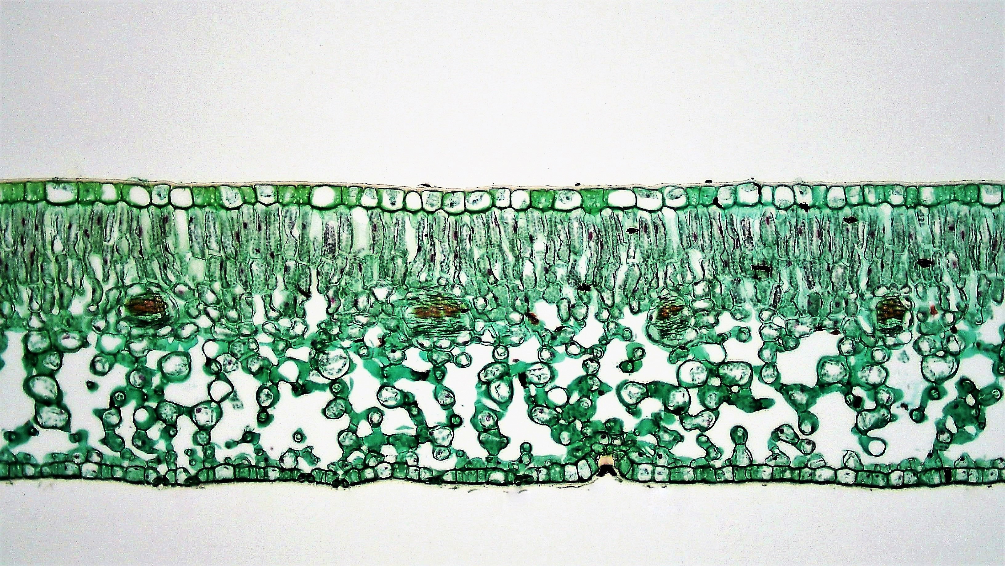
Transverse section of Ligustrum (privet) leaf showing typical mesophytic dicot anatomy: upper and lower epidermis with cuticle, palisade mesophyll layer, spongy mesophyll with intercellular air spaces, and vascular bundles.
Classic mesophytic dicot leaf anatomy — Ligustrum (privet)
Wikimedia Commons — CC BY-SA 3.0
Transverse section of Ficus leaf showing xerophytic adaptations: thick cuticle, multi-layered epidermis with cystoliths, compact palisade mesophyll, reduced spongy mesophyll, and prominent midrib vascular bundle.
Xerophytic dicot leaf anatomy — Ficus with cystoliths
Wikimedia Commons — CC BY-SA 3.0
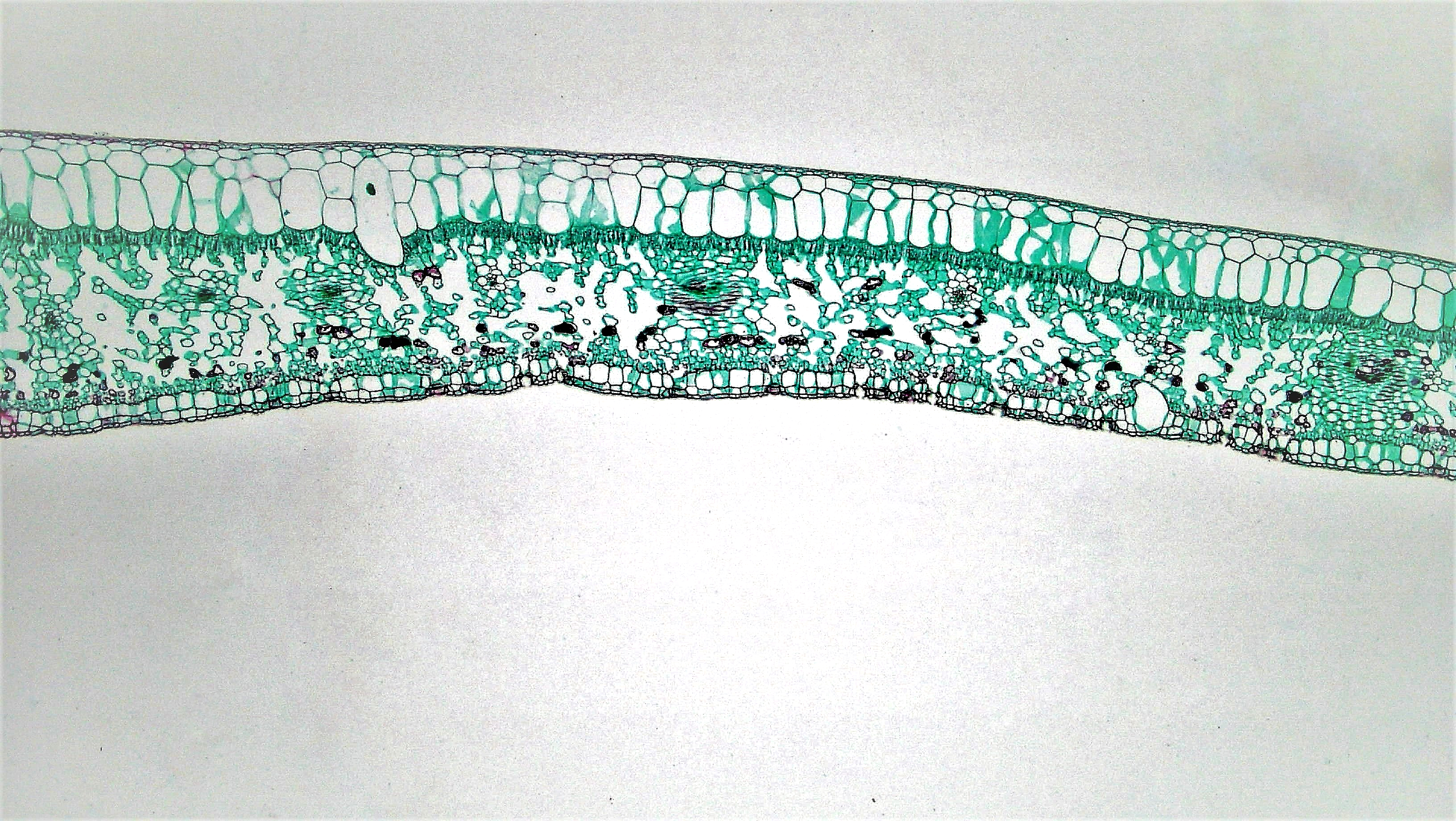
Higher magnification view of Ligustrum leaf mesophyll showing distinct palisade mesophyll (elongated cells with chloroplasts) and spongy mesophyll (irregular cells with large intercellular spaces).
Mesophyll detail — palisade and spongy layer comparison
Wikimedia Commons — CC BY-SA 3.0
Compatible Microscopes
| Model | Manufacturer | Type | Magnification Range | NA Max | Resolution |
|---|---|---|---|---|---|
| VHX-7000 | Keyence | Digital Microscope | 0.1–6000× | — | 100 nm |
| DM6 B | Leica Microsystems | Upright Optical | 1.25–100× | 1.4 | 200 nm |
| S9i | Leica Microsystems | Stereo | 6.1–55× | — | — nm |
| Eclipse Ni | Nikon | Upright Optical | 2–100× | 1.4 | 200 nm |
| SMZ25 | Nikon | Stereo | 0.63–15.75× | — | — nm |
| BX53 | Olympus (Evident) | Upright Optical | 2–100× | 1.4 | 200 nm |
| SZX16 | Olympus (Evident) | Stereo | 0.7–11.5× | 0.3 | — nm |
| Axio Imager 2 | Zeiss | Upright Optical | 1.25–100× | 1.4 | 200 nm |
| Primostar 3 | Zeiss | Upright Optical | 4–100× | 1.25 | 350 nm |
| Stemi 508 | Zeiss | Stereo | 6.3–50× | — | — nm |
Edit Image Metadata
AI Generate Image — Plant Leaf Cross-Section
Fill in the imaging criteria below. A detailed prompt will be built from your selections and sent to OpenAI to generate a realistic microscopy image.