Urinalysis Sediment
Urinalysis Sediment
Clinical ChemistryCentrifuged urine sediment showing casts, crystals, and cells
Drag & drop images here to add to this specimen
or click Upload Image aboveRelease to upload
Image Library
3 images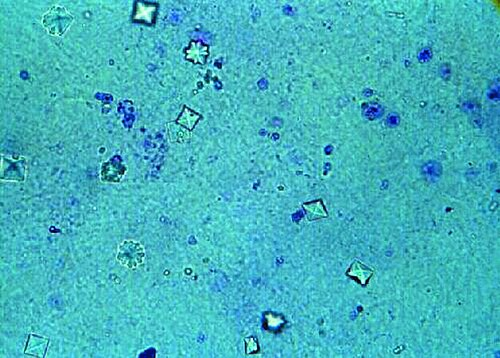
Unstained urine sediment showing characteristic envelope-shaped (bipyramidal) calcium oxalate dihydrate crystals. These are the most common crystals found in acidic urine. Red blood cells are visible in the background.
Calcium oxalate dihydrate — classic envelope morphology, clinically significant
Wikimedia Commons — CC BY-SA 3.0
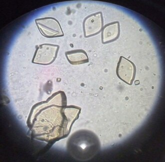
Unstained urine sediment showing uric acid crystals with characteristic rhomboid or rosette morphology. Uric acid crystals are found in acidic urine and may be associated with gout or high purine diet.
Uric acid crystals — rhomboid morphology, acidic urine, may indicate gout
Wikimedia Commons — CC BY-SA 3.0
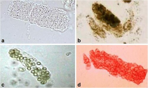
Urine sediment microscopy showing multiple cast types including renal tubular epithelial (RTE) casts, muddy brown granular casts, WBC casts, and RBC casts. These cylindrical structures form in the renal tubules and indicate kidney pathology.
Multiple cast types — RTE, granular, WBC, RBC casts indicating renal disease
Wikimedia Commons — CC BY-SA 3.0
Compatible Microscopes
| Model | Manufacturer | Type | Magnification Range | NA Max | Resolution |
|---|---|---|---|---|---|
| DM6 B | Leica Microsystems | Upright Optical | 1.25–100× | 1.4 | 200 nm |
| DMi8 | Leica Microsystems | Inverted Optical | 2.5–100× | 1.47 | 185 nm |
| Eclipse Ti2 | Nikon | Inverted Optical | 2–100× | 1.45 | 190 nm |
| Eclipse Ni | Nikon | Upright Optical | 2–100× | 1.4 | 200 nm |
| BX53 | Olympus (Evident) | Upright Optical | 2–100× | 1.4 | 200 nm |
| IX83 | Olympus (Evident) | Inverted Optical | 2–100× | 1.4 | 200 nm |
| Axio Observer 7 | Zeiss | Inverted Optical | 5–100× | 1.4 | 200 nm |
| Axio Imager 2 | Zeiss | Upright Optical | 1.25–100× | 1.4 | 200 nm |
| Primostar 3 | Zeiss | Upright Optical | 4–100× | 1.25 | 350 nm |
Edit Image Metadata
AI Generate Image — Urinalysis Sediment
Fill in the imaging criteria below. A detailed prompt will be built from your selections and sent to OpenAI to generate a realistic microscopy image.