Brain Tissue (Neurons)
Brain Tissue (Neurons)
NeuroscienceGolgi-stained brain tissue section showing neuronal morphology
Drag & drop images here to add to this specimen
or click Upload Image aboveRelease to upload
Image Library
3 images
Golgi-stained hippocampal pyramidal neuron showing the cell body (soma), apical dendrite extending upward, and basal dendrites radiating from the soma. Dendritic spines are visible along the branches.
Golgi silver impregnation — classic neuron morphology preparation
Wikimedia Commons — CC BY-SA 3.0

Golgi silver-stained cortical pyramidal neuron from the cerebral cortex. The characteristic triangular cell body, prominent apical dendrite, and extensive dendritic arbor are clearly visible against the unstained background.
Golgi method — random sparse staining reveals individual neuron morphology
Wikimedia Commons — Public Domain
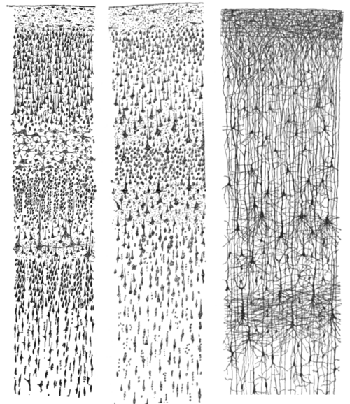
Histological preparation of cerebral cortex tissue showing layers of neurons. Different neuronal types are visible including stellate and pyramidal cells across cortical layers.
Cortical architecture with visible laminar organization
Wikimedia Commons — Public Domain
Compatible Microscopes
| Model | Manufacturer | Type | Magnification Range | NA Max | Resolution |
|---|---|---|---|---|---|
| DM6 B | Leica Microsystems | Upright Optical | 1.25–100× | 1.4 | 200 nm |
| DMi8 | Leica Microsystems | Inverted Optical | 2.5–100× | 1.47 | 185 nm |
| STELLARIS 5 | Leica Microsystems | Confocal | 5–100× | — | 120 nm |
| Eclipse Ti2 | Nikon | Inverted Optical | 2–100× | 1.45 | 190 nm |
| Eclipse Ni | Nikon | Upright Optical | 2–100× | 1.4 | 200 nm |
| A1R HD25 | Nikon | Confocal | 4–100× | — | 140 nm |
| BX53 | Olympus (Evident) | Upright Optical | 2–100× | 1.4 | 200 nm |
| IX83 | Olympus (Evident) | Inverted Optical | 2–100× | 1.4 | 200 nm |
| FV4000 | Olympus (Evident) | Confocal | 4–100× | — | 120 nm |
| Axio Observer 7 | Zeiss | Inverted Optical | 5–100× | 1.4 | 200 nm |
| Axio Imager 2 | Zeiss | Upright Optical | 1.25–100× | 1.4 | 200 nm |
| LSM 980 | Zeiss | Confocal | 5–63× | — | 120 nm |
| Primostar 3 | Zeiss | Upright Optical | 4–100× | 1.25 | 350 nm |
Edit Image Metadata
AI Generate Image — Brain Tissue (Neurons)
Fill in the imaging criteria below. A detailed prompt will be built from your selections and sent to OpenAI to generate a realistic microscopy image.