HeLa Cell Culture
HeLa Cell Culture
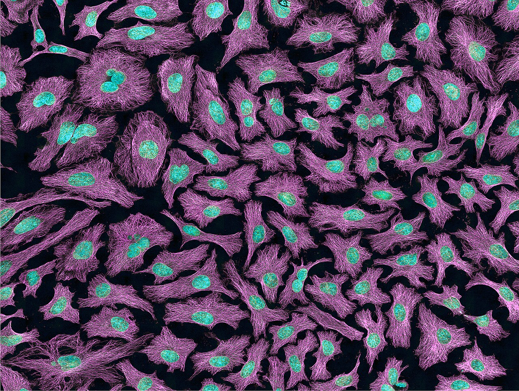
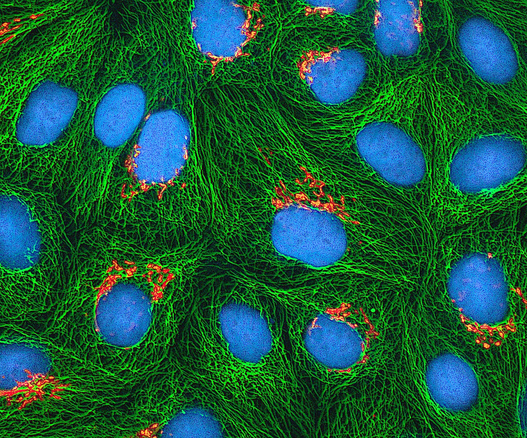
Cell BiologyCultured HeLa cells (cervical adenocarcinoma) in monolayer
Drag & drop images here to add to this specimen
or click Upload Image aboveRelease to upload
Image Library
3 images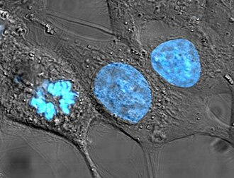
HeLa cervical carcinoma cells stained with Hoechst 33258 nuclear dye. Nuclei fluoresce bright blue against a dark background, revealing nuclear morphology, mitotic figures, and chromatin condensation patterns.
Hoechst 33258 — DNA-binding fluorescent dye highlighting nuclei
Wikimedia Commons — CC BY-SA 3.0
Phase contrast microscopy image of HeLa cells in culture showing the characteristic epithelial-like monolayer morphology. Cells are adherent with visible cell boundaries and refractile cytoplasm.
Phase contrast — unstained living cells in culture flask
Wikimedia Commons — CC BY 3.0
Inverted microscopy image of HeLa cells growing in monolayer culture. The cells show typical epithelioid morphology with irregular polygonal shapes and prominent nuclei visible in phase contrast.
Standard HeLa cell line morphology in culture
Wikimedia Commons — CC BY 3.0
Compatible Microscopes
| Model | Manufacturer | Type | Magnification Range | NA Max | Resolution |
|---|---|---|---|---|---|
| BZ-X810 | Keyence | Fluorescence All In One | 2–100× | 1.45 | 190 nm |
| DMi8 | Leica Microsystems | Inverted Optical | 2.5–100× | 1.47 | 185 nm |
| STELLARIS 5 | Leica Microsystems | Confocal | 5–100× | — | 120 nm |
| Eclipse Ti2 | Nikon | Inverted Optical | 2–100× | 1.45 | 190 nm |
| A1R HD25 | Nikon | Confocal | 4–100× | — | 140 nm |
| IX83 | Olympus (Evident) | Inverted Optical | 2–100× | 1.4 | 200 nm |
| FV4000 | Olympus (Evident) | Confocal | 4–100× | — | 120 nm |
| Axio Observer 7 | Zeiss | Inverted Optical | 5–100× | 1.4 | 200 nm |
| LSM 980 | Zeiss | Confocal | 5–63× | — | 120 nm |
Edit Image Metadata
AI Generate Image — HeLa Cell Culture
Fill in the imaging criteria below. A detailed prompt will be built from your selections and sent to OpenAI to generate a realistic microscopy image.