Human Genome
Human Genome
CytogeneticsHuman chromosome preparations for cytogenetic analysis including G-banded karyotypes, metaphase spreads, and FISH (fluorescence in situ hybridization)
Drag & drop images here to add to this specimen
or click Upload Image aboveRelease to upload
Image Library
3 images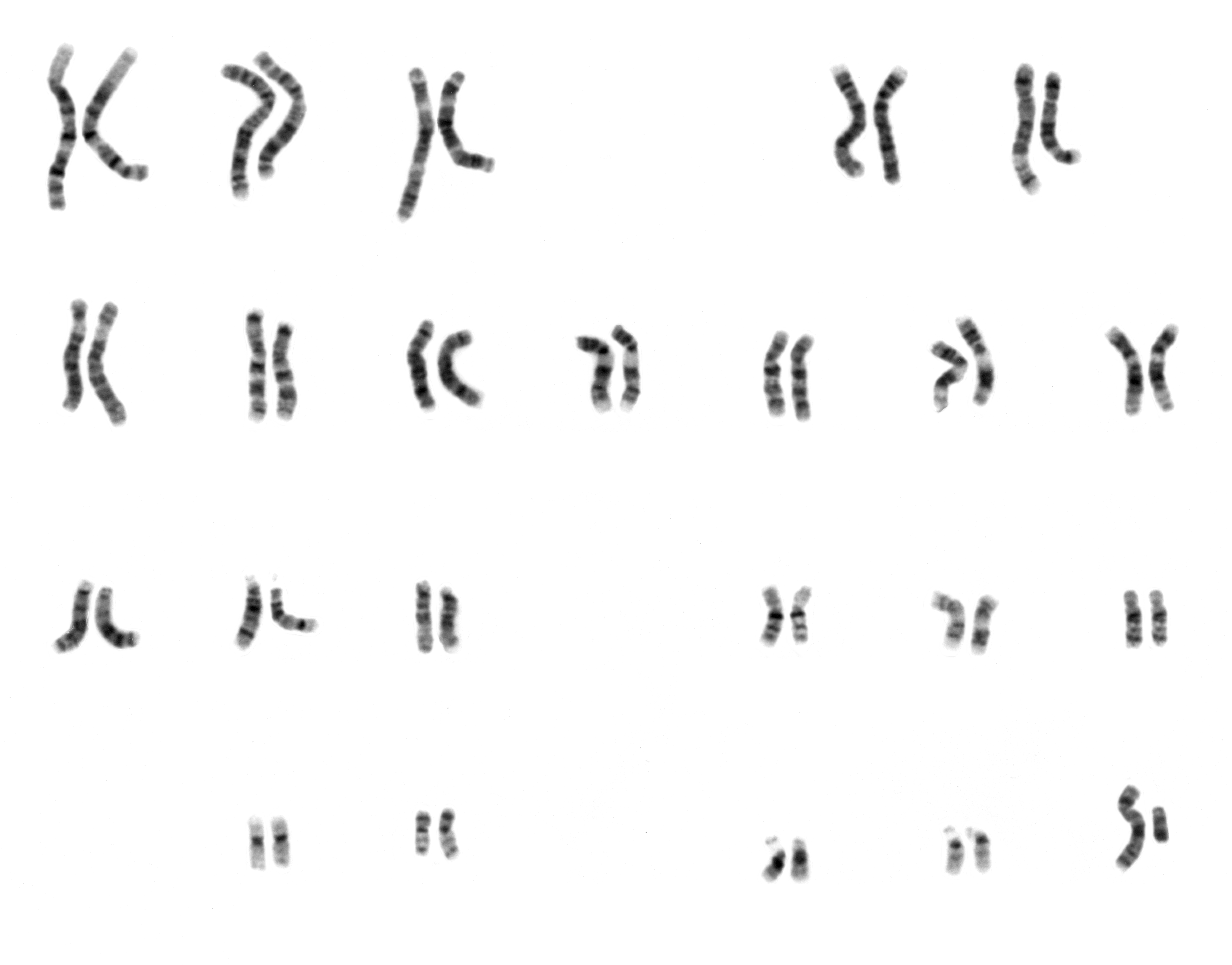
Complete G-banded karyotype of a human male (46,XY) showing all 22 pairs of autosomes and the X and Y sex chromosomes. Each chromosome is arranged by size and banding pattern, with characteristic Giemsa dark and light bands visible for cytogenetic identification and analysis.
G-banded karyotype 46,XY — NHGRI reference standard
Wikimedia Commons — Public Domain (NHGRI)
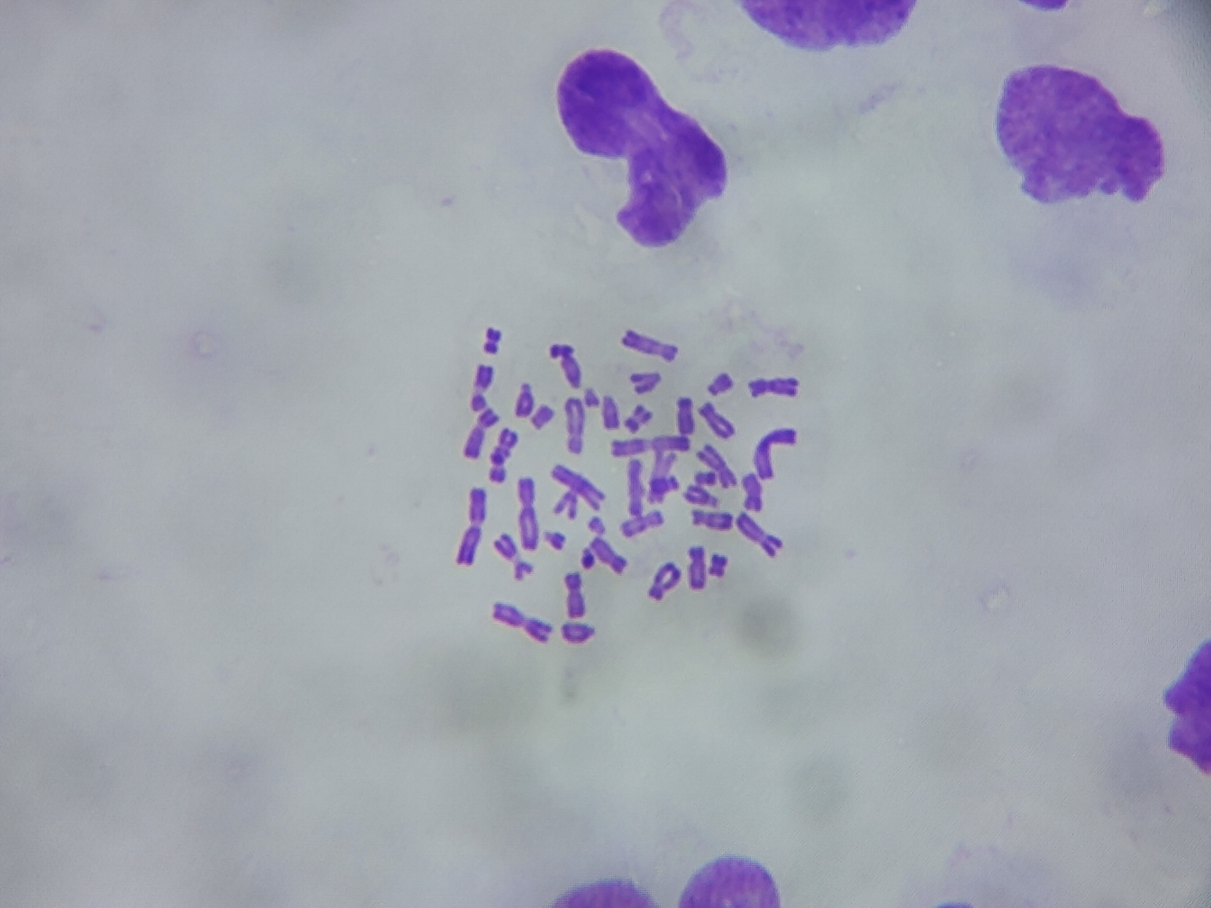
Brightfield microscopy image of a human metaphase chromosome spread preparation. Individual chromosomes are visible as distinct condensed structures arrested during cell division, suitable for cytogenetic analysis, counting, and banding studies.
Metaphase spread — chromosome counting and morphology
Wikimedia Commons — CC BY 4.0
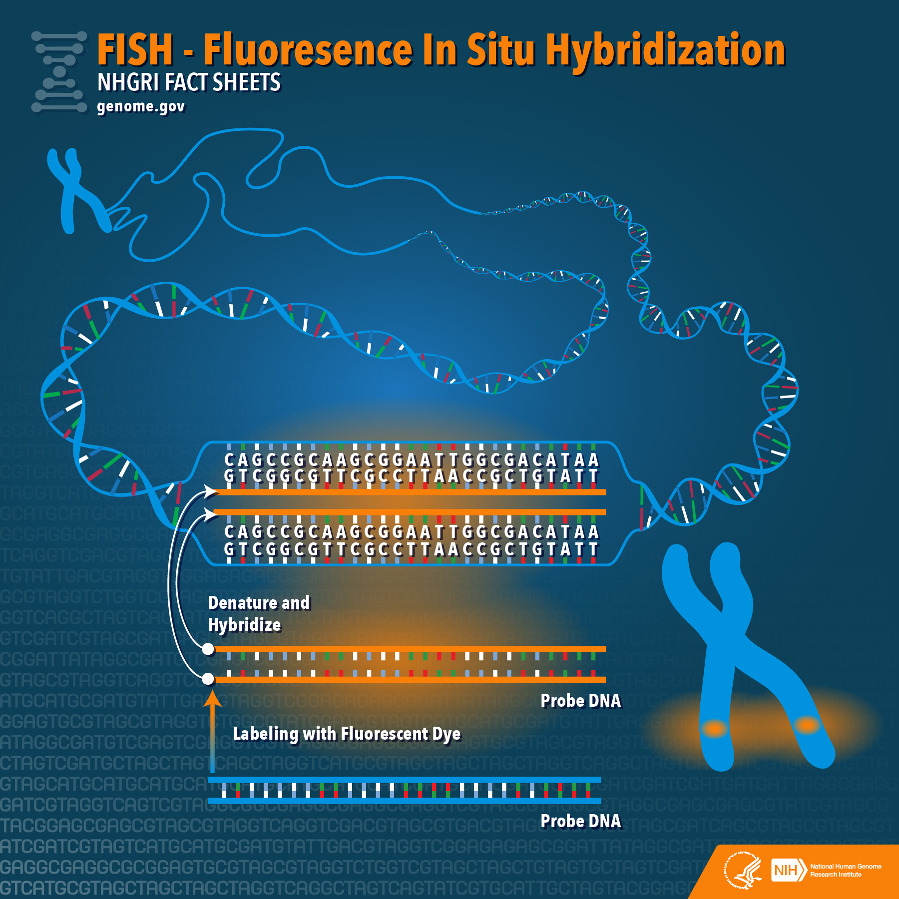
Fluorescence in situ hybridization (FISH) image showing fluorescently labeled DNA probes hybridized to specific chromosomal regions. FISH enables visualization and mapping of genetic material within individual cells, identifying specific genes or chromosomal segments through complementary fluorescent probe binding.
FISH — gene mapping with fluorescent DNA probes
Wikimedia Commons — CC BY 2.0 (NHGRI)
Compatible Microscopes
| Model | Manufacturer | Type | Magnification Range | NA Max | Resolution |
|---|---|---|---|---|---|
| BZ-X810 | Keyence | Fluorescence All In One | 2–100× | 1.45 | 190 nm |
| VHX-7000 | Keyence | Digital Microscope | 0.1–6000× | — | 100 nm |
| DM6 B | Leica Microsystems | Upright Optical | 1.25–100× | 1.4 | 200 nm |
| STELLARIS 5 | Leica Microsystems | Confocal | 5–100× | — | 120 nm |
| Eclipse Ni | Nikon | Upright Optical | 2–100× | 1.4 | 200 nm |
| A1R HD25 | Nikon | Confocal | 4–100× | — | 140 nm |
| BX53 | Olympus (Evident) | Upright Optical | 2–100× | 1.4 | 200 nm |
| FV4000 | Olympus (Evident) | Confocal | 4–100× | — | 120 nm |
| Axio Imager 2 | Zeiss | Upright Optical | 1.25–100× | 1.4 | 200 nm |
| LSM 980 | Zeiss | Confocal | 5–63× | — | 120 nm |
| Primostar 3 | Zeiss | Upright Optical | 4–100× | 1.25 | 350 nm |
Edit Image Metadata
AI Generate Image — Human Genome
Fill in the imaging criteria below. A detailed prompt will be built from your selections and sent to OpenAI to generate a realistic microscopy image.