Virus Particles (Negative Stain)
Virus Particles (Negative Stain)
VirologyNegatively stained virus particles on carbon-coated copper grid
Drag & drop images here to add to this specimen
or click Upload Image aboveRelease to upload
Image Library
5 images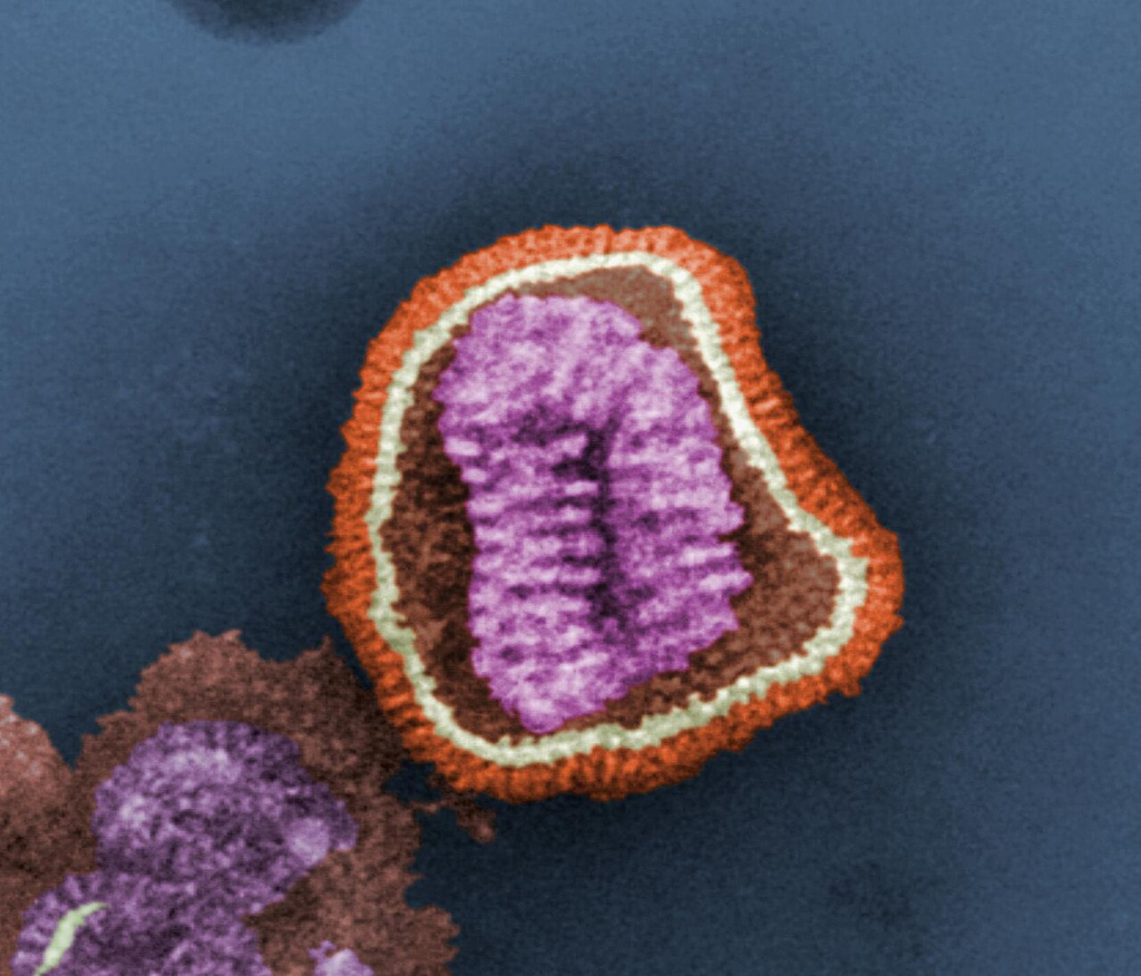
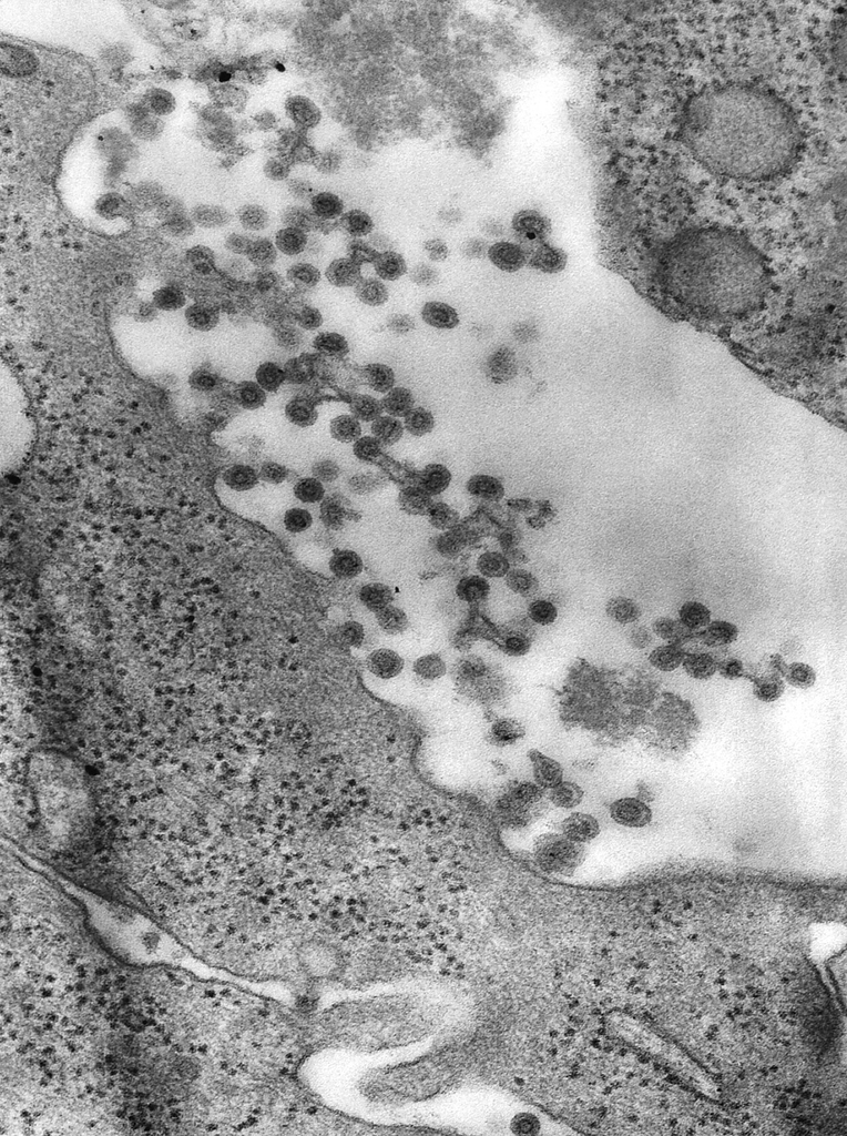
Transmission electron micrograph of influenza virus particles (approximately 80–120 nm diameter) showing the characteristic spherical/pleomorphic morphology with surface projections (hemagglutinin and neuraminidase spikes). Negatively stained with phosphotungstic acid.
Negative stain TEM — influenza virion with surface glycoprotein spikes
Wikimedia Commons — Public Domain (CDC)
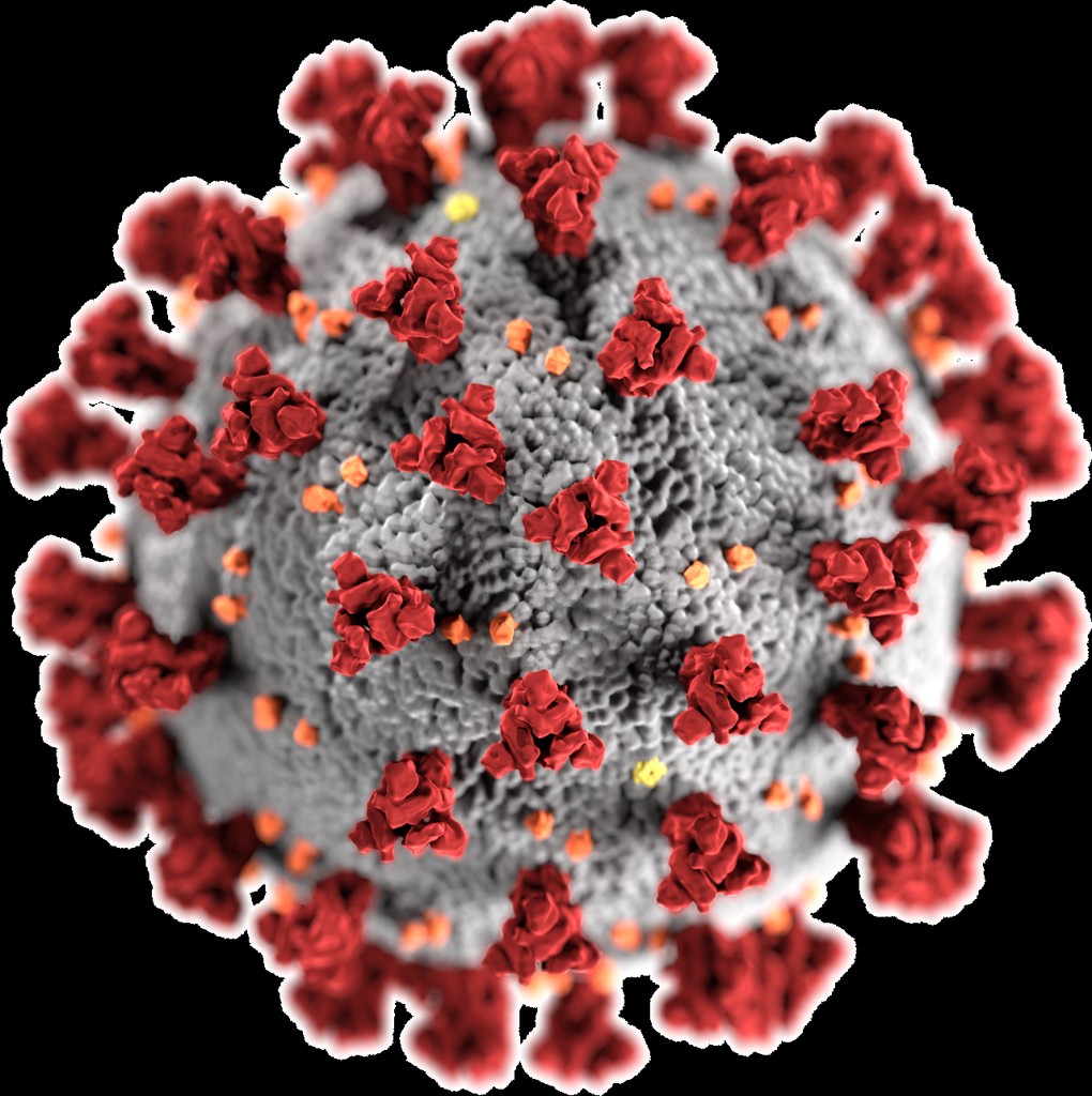
Transmission electron micrograph of SARS-CoV-2 virus particles showing the characteristic crown-like (corona) spike proteins projecting from the viral envelope. Particles are approximately 100–120 nm in diameter with a helical nucleocapsid inside the lipid bilayer envelope.
SARS-CoV-2 — envelope, spike (S) proteins, nucleocapsid visible
Wikimedia Commons — Public Domain (NIAID)
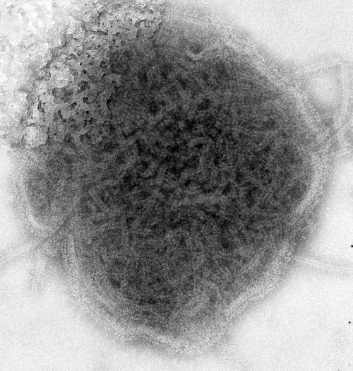
Transmission electron micrograph of mumps virus particles negatively stained with phosphotungstic acid. The pleomorphic enveloped virions (100–600 nm) show surface glycoprotein projections and internal ribonucleoprotein.
Negative stain TEM — mumps paramyxovirus, pleomorphic enveloped virions
Wikimedia Commons — Public Domain (CDC)
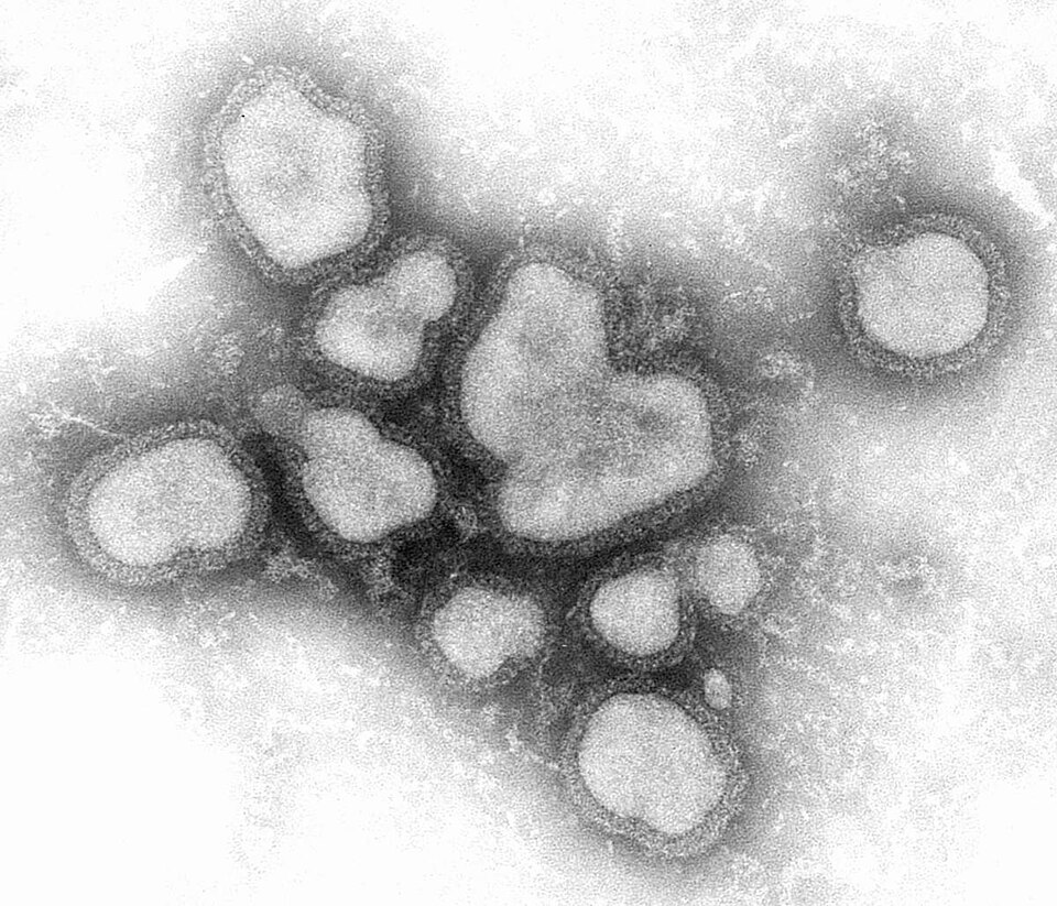
Negatively stained TEM image of influenza A virus particles showing characteristic spherical and filamentous morphologies. Surface spikes (hemagglutinin and neuraminidase) project from the viral envelope.
Negative stain — influenza A virion with HA and NA surface glycoproteins
Wikimedia Commons — Public Domain (CDC)
Transmission electron micrograph of rubella virus particles showing the enveloped spherical virions (approximately 60–70 nm diameter). The lipid envelope with embedded glycoprotein spikes surrounds the nucleocapsid core.
Rubella togavirus — enveloped spherical virion, ~60-70 nm
Wikimedia Commons — Public Domain (CDC)
Compatible Microscopes
| Model | Manufacturer | Type | Magnification Range | NA Max | Resolution |
|---|---|---|---|---|---|
| HT7800 | Hitachi High-Tech | Tem | 200–600000× | — | — nm |
| JEM-F200 | JEOL | Tem | 50–1500000× | — | — nm |
| JEM-ARM300F2 | JEOL | Stem | 100–150000000× | — | — nm |
| Titan Krios G4 | Thermo Fisher Scientific | Cryo Tem | 640–1050000× | — | — nm |
Edit Image Metadata
AI Generate Image — Virus Particles (Negative Stain)
Fill in the imaging criteria below. A detailed prompt will be built from your selections and sent to OpenAI to generate a realistic microscopy image.