Dental — Periapical View
Dental — Periapical View
DentalPeriapical radiograph phantom showing 2-3 teeth with roots, periodontal ligament, alveolar bone, and surrounding structures. Standard intraoral dental X-ray view
Image Library
Periapical radiograph showing tooth and surrounding structures
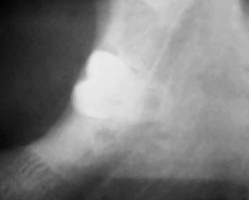
https://commons.wikimedia.org/wiki/File:Radiograph%20of%20impacted%20wisdom%20tooth%20near%20nerve.jpg
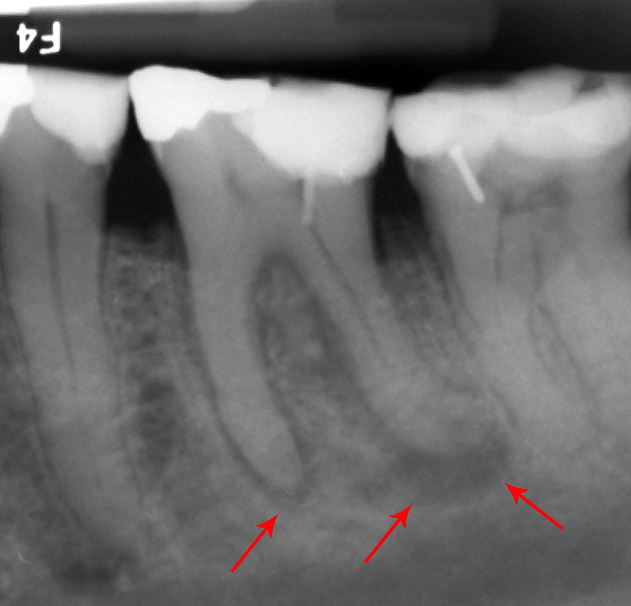
Periapical radiograph showing teeth roots and alveolar bone
https://commons.wikimedia.org/wiki/File:Abscessed%20tooth%20periapical%20radiograph.jpg
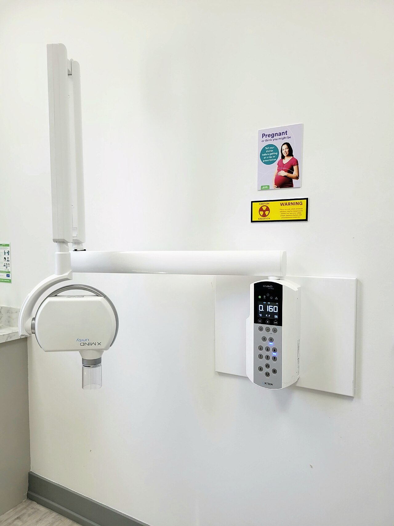
Intraoral dental radiograph showing dental anatomy
https://commons.wikimedia.org/wiki/File:X-Mind%20Unity%20dental%20X-ray%20generator%20for%20Intraoral%20Imaging.jpg
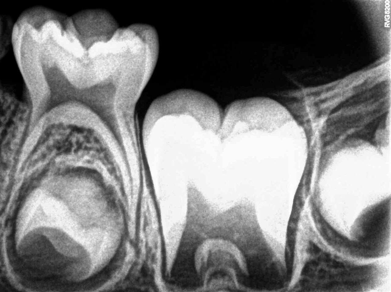
Periapical radiograph showing tooth and surrounding structures
https://commons.wikimedia.org/wiki/File:Intraoral%20Periapical%20Radiograph%20%28IOPA%29%20showing%20Deciduous%28Milky%20or%20Primary%29%20Tooth%2075%20and%20developing%20crown%20of%20Permanent%20or%20Secondary%20Teeth%2035%2C%2036%20and%2037.jpg
Tissue Structures
| Structure | Density (HU) | T1 (ms) | T2 (ms) | Echo | Rel. Size | |
|---|---|---|---|---|---|---|
| Enamel | 2500 | 0 | 0 | 0.0 | 12% | |
| Dentin | 1600 | 0 | 0 | 0.0 | 18% | |
| Pulp Chamber | 20 | 2000 | 200 | 0.0 | 3% | |
| Root Canal | 20 | 2000 | 200 | 0.0 | 2% | |
| Cementum | 1200 | 0 | 0 | 0.0 | 4% | |
| Periodontal Ligament | 30 | 1200 | 60 | 0.0 | 3% | |
| Alveolar Bone | 800 | 350 | 3 | 0.9 | 20% | |
| Cortical Plate | 1400 | 300 | 1 | 0.95 | 6% | |
| Cancellous Bone | 300 | 400 | 50 | 0.7 | 15% | |
| Gingiva | 30 | 800 | 45 | 0.3 | 5% |
Compatible Imaging Instruments
| Model | Manufacturer | Modality |
|---|---|---|
| Aquilion ONE PRISM | Canon Medical Systems | CT |
| SCENARIA View | Fujifilm Healthcare | CT |
| Revolution Apex | GE HealthCare | CT |
| Spectral CT 7500 | Philips Healthcare | CT |
| SOMATOM Force | Siemens Healthineers | CT |
Edit Image Metadata
AI Generate Image — Dental — Periapical View
Fill in the imaging criteria below. A prompt will be built and sent to OpenAI to generate a realistic medical image.