Gold Nanoparticles
Gold Nanoparticles
NanotechnologyGold nanoparticles (50 nm) on carbon grid for TEM imaging
Drag & drop images here to add to this specimen
or click Upload Image aboveRelease to upload
Image Library
3 images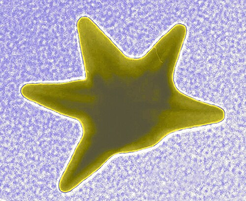
TEM image of gold nanostars showing their characteristic branched morphology with multiple pointed tips extending from a central core. These anisotropic nanoparticles exhibit unique optical properties due to localized surface plasmon resonance at the tips.
Gold nanostars — branched morphology, LSPR-active tips
Wikimedia Commons — CC BY-SA 3.0
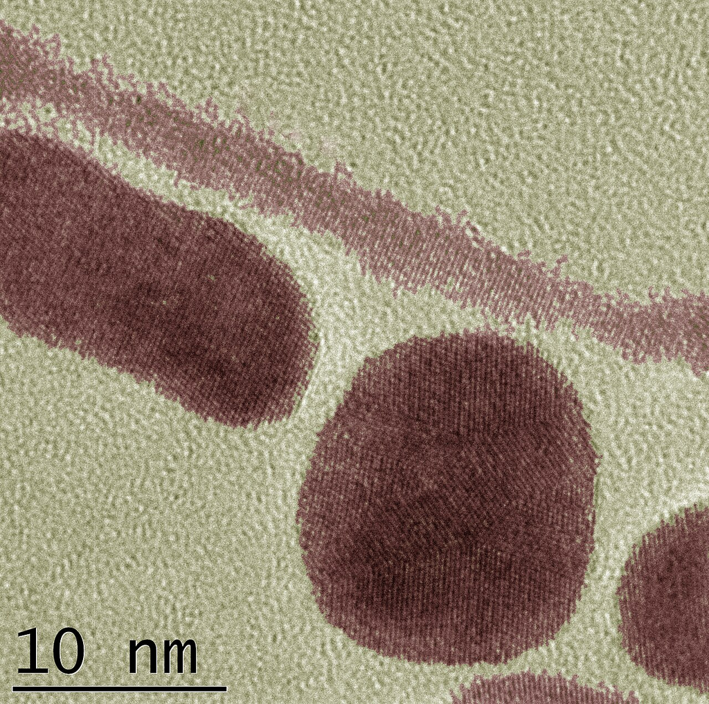
Transmission electron micrograph of gold nanoparticles and nanorods deposited on a carbon-coated copper grid. Spherical particles and elongated rod-shaped nanostructures are visible with characteristic electron-dense contrast.
TEM bright-field — AuNPs and AuNRs on carbon support film
Wikimedia Commons — CC BY-SA 3.0
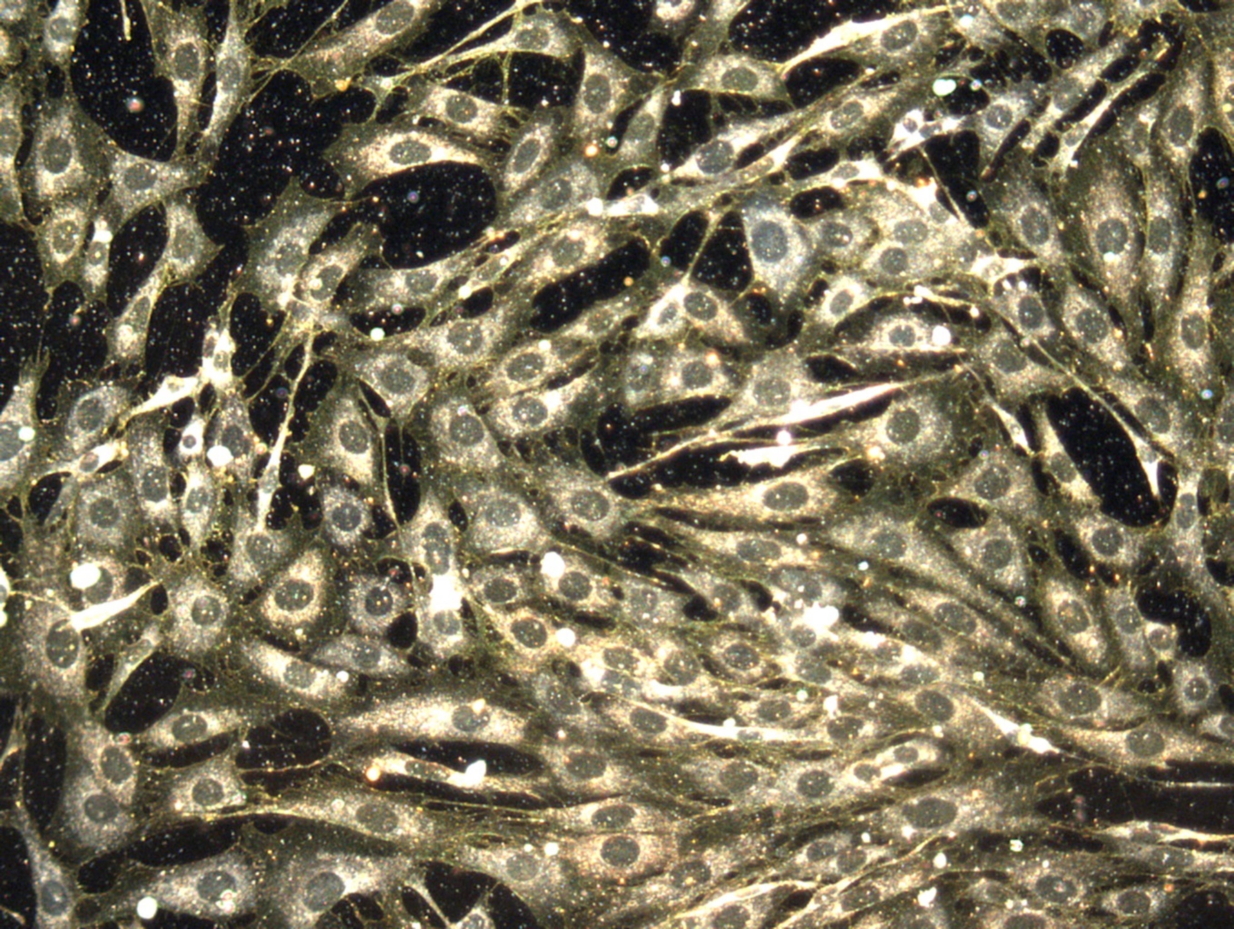
Transmission electron micrograph showing gold nanoparticles internalized within the cytosol of a cell. The electron-dense gold particles appear as dark spherical dots against the cellular ultrastructure, demonstrating nanoparticle uptake and distribution at the subcellular level.
TEM — intracellular gold nanoparticle distribution
Wikimedia Commons — CC BY-SA 3.0
Compatible Microscopes
| Model | Manufacturer | Type | Magnification Range | NA Max | Resolution |
|---|---|---|---|---|---|
| HT7800 | Hitachi High-Tech | Tem | 200–600000× | — | — nm |
| Regulus 8200 | Hitachi High-Tech | Fe Sem | 20–2000000× | — | 0.6 nm |
| JEM-F200 | JEOL | Tem | 50–1500000× | — | — nm |
| JEM-ARM300F2 | JEOL | Stem | 100–150000000× | — | — nm |
| JSM-IT800 | JEOL | Sem | 5–1000000× | — | 0.7 nm |
| JSM-7900F | JEOL | Fe Sem | 25–1000000× | — | 0.5 nm |
| Titan Krios G4 | Thermo Fisher Scientific | Cryo Tem | 640–1050000× | — | — nm |
| Talos F200X G2 | Thermo Fisher Scientific | Analytical Tem Stem | 25–2050000× | — | — nm |
| Apreo 2 | Thermo Fisher Scientific | Fe Sem | 13–2000000× | — | 0.7 nm |
Edit Image Metadata
AI Generate Image — Gold Nanoparticles
Fill in the imaging criteria below. A detailed prompt will be built from your selections and sent to OpenAI to generate a realistic microscopy image.